8S7C

Ternary Complex of Cachd1, FZD5 and LRP6


Experimental Data Snapshot

  • Method: X-RAY DIFFRACTION
  • Resolution: 4.70 Å
  • R-Value Free: 0.261 
  • R-Value Work: 0.206 
  • R-Value Observed: 0.209 

wwPDB Validation   3D Report Full Report


Ligand Structure Quality Assessment 


This is version 1.1 of the entry. See complete history


Literature

Cachd1 interacts with Wnt receptors and regulates neuronal asymmetry in the zebrafish brain.

Powell, G.T.Faro, A.Zhao, Y.Stickney, H.Novellasdemunt, L.Henriques, P.Gestri, G.Redhouse White, E.Ren, J.Lu, W.Young, R.M.Hawkins, T.A.Cavodeassi, F.Schwarz, Q.Dreosti, E.Raible, D.W.Li, V.S.W.Wright, G.J.Jones, E.Y.Wilson, S.W.

(2024) Science 384: 573-579

  • DOI: https://doi.org/10.1126/science.ade6970
  • Primary Citation of Related Structures:  
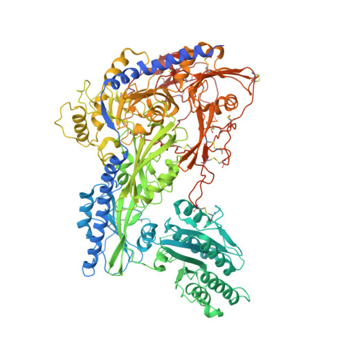
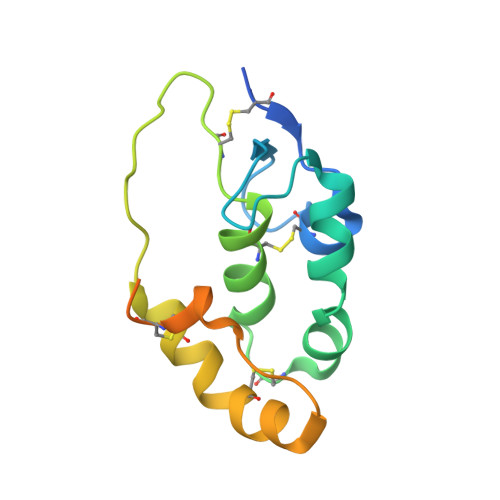
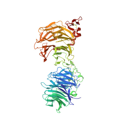
    8S7C

  • PubMed Abstract: 

    Neurons on the left and right sides of the nervous system often show asymmetric properties, but how such differences arise is poorly understood. Genetic screening in zebrafish revealed that loss of function of the transmembrane protein Cachd1 resulted in right-sided habenula neurons adopting left-sided identity. Cachd1 is expressed in neuronal progenitors, functions downstream of asymmetric environmental signals, and influences timing of the normally asymmetric patterns of neurogenesis. Biochemical and structural analyses demonstrated that Cachd1 can bind simultaneously to Lrp6 and Frizzled family Wnt co-receptors. Consistent with this, lrp6 mutant zebrafish lose asymmetry in the habenulae, and epistasis experiments support a role for Cachd1 in modulating Wnt pathway activity in the brain. These studies identify Cachd1 as a conserved Wnt receptor-interacting protein that regulates lateralized neuronal identity in the zebrafish brain.


  • Organizational Affiliation

    Cell and Developmental Biology, Division of Biosciences, University College London, London WC1E 6BT, UK.


Macromolecules
Find similar proteins by:  (by identity cutoff)  |  3D Structure
Entity ID: 1
MoleculeChains Sequence LengthOrganismDetailsImage
VWFA and cache domain-containing protein 1
A, D, G
1,102Mus musculusMutation(s): 0 
Gene Names: Cachd1Kiaa1573Vwcd1
UniProt & NIH Common Fund Data Resources
Find proteins for Q6PDJ1 (Mus musculus)
Explore Q6PDJ1 
Go to UniProtKB:  Q6PDJ1
IMPC:  MGI:2444177
Entity Groups  
Sequence Clusters30% Identity50% Identity70% Identity90% Identity95% Identity100% Identity
UniProt GroupQ6PDJ1
Sequence Annotations
Expand
  • Reference Sequence
Find similar proteins by:  (by identity cutoff)  |  3D Structure
Entity ID: 2
MoleculeChains Sequence LengthOrganismDetailsImage
Frizzled-5
B, E, H
140Mus musculusMutation(s): 0 
Gene Names: Fzd5
UniProt & NIH Common Fund Data Resources
Find proteins for Q9EQD0 (Mus musculus)
Explore Q9EQD0 
Go to UniProtKB:  Q9EQD0
IMPC:  MGI:108571
Entity Groups  
Sequence Clusters30% Identity50% Identity70% Identity90% Identity95% Identity100% Identity
UniProt GroupQ9EQD0
Sequence Annotations
Expand
  • Reference Sequence
Find similar proteins by:  (by identity cutoff)  |  3D Structure
Entity ID: 3
MoleculeChains Sequence LengthOrganismDetailsImage
Low-density lipoprotein receptor-related protein 6
C, F, I
628Homo sapiensMutation(s): 0 
Gene Names: LRP6
UniProt & NIH Common Fund Data Resources
Find proteins for O75581 (Homo sapiens)
Explore O75581 
Go to UniProtKB:  O75581
PHAROS:  O75581
GTEx:  ENSG00000070018 
Entity Groups  
Sequence Clusters30% Identity50% Identity70% Identity90% Identity95% Identity100% Identity
UniProt GroupO75581
Sequence Annotations
Expand
  • Reference Sequence
Oligosaccharides

Help

Entity ID: 4
MoleculeChains Length2D Diagram Glycosylation3D Interactions
2-acetamido-2-deoxy-beta-D-glucopyranose-(1-4)-2-acetamido-2-deoxy-beta-D-glucopyranose
J, K
2N-Glycosylation
Glycosylation Resources
GlyTouCan:  G42666HT
GlyCosmos:  G42666HT
GlyGen:  G42666HT
Small Molecules
Ligands 1 Unique
IDChains Name / Formula / InChI Key2D Diagram3D Interactions
NAG (Subject of Investigation/LOI)
Query on NAG

Download Ideal Coordinates CCD File 
AA [auth F]
BA [auth F]
CA [auth F]
DA [auth F]
EA [auth F]
AA [auth F],
BA [auth F],
CA [auth F],
DA [auth F],
EA [auth F],
FA [auth G],
GA [auth G],
HA [auth G],
IA [auth G],
JA [auth G],
KA [auth G],
L [auth A],
LA [auth H],
M [auth A],
MA [auth I],
N [auth A],
NA [auth I],
O [auth A],
OA [auth I],
P [auth A],
Q [auth C],
R [auth C],
S [auth C],
T [auth C],
U [auth D],
V [auth D],
W [auth D],
X [auth D],
Y [auth D],
Z [auth E]
2-acetamido-2-deoxy-beta-D-glucopyranose
C8 H15 N O6
OVRNDRQMDRJTHS-FMDGEEDCSA-N
Experimental Data & Validation

Experimental Data

  • Method: X-RAY DIFFRACTION
  • Resolution: 4.70 Å
  • R-Value Free: 0.261 
  • R-Value Work: 0.206 
  • R-Value Observed: 0.209 
  • Space Group: C 1 2 1
Unit Cell:
Length ( Å )Angle ( ˚ )
a = 283.697α = 90
b = 198.236β = 128.07
c = 218.824γ = 90
Software Package:
Software NamePurpose
PHENIXrefinement
xia2data scaling
xia2data reduction
PHENIXphasing

Structure Validation

View Full Validation Report


Ligand Structure Quality Assessment 


Entry History & Funding Information

Deposition Data


Funding OrganizationLocationGrant Number
Cancer Research UKUnited KingdomC375/A17721
Wellcome TrustUnited Kingdom223133/Z/21/Z

Revision History  (Full details and data files)

  • Version 1.0: 2024-04-24
    Type: Initial release
  • Version 1.1: 2024-05-15
    Changes: Database references