8OZ7

Abortive infection DNA polymerase AbiA from Lactococcus lactis


Experimental Data Snapshot

  • Method: X-RAY DIFFRACTION
  • Resolution: 2.75 Å
  • R-Value Free: 0.264 
  • R-Value Work: 0.220 
  • R-Value Observed: 0.221 

wwPDB Validation   3D Report Full Report


This is version 1.2 of the entry. See complete history


Literature

Structure-functional characterization of Lactococcus AbiA phage defense system.

Gapinska, M.Zajko, W.Skowronek, K.Figiel, M.Krawczyk, P.S.Egorov, A.A.Dziembowski, A.Johansson, M.J.O.Nowotny, M.

(2024) Nucleic Acids Res 52: 4723-4738

  • DOI: https://doi.org/10.1093/nar/gkae230
  • Primary Citation of Related Structures:  
    8OZ7

  • PubMed Abstract: 

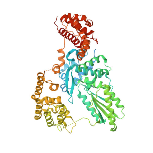
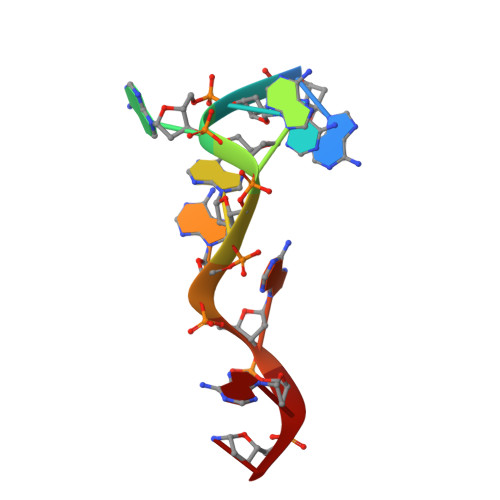
    Bacterial reverse transcriptases (RTs) are a large and diverse enzyme family. AbiA, AbiK and Abi-P2 are abortive infection system (Abi) RTs that mediate defense against bacteriophages. What sets Abi RTs apart from other RT enzymes is their ability to synthesize long DNA products of random sequences in a template- and primer-independent manner. Structures of AbiK and Abi-P2 representatives have recently been determined, but there are no structural data available for AbiA. Here, we report the crystal structure of Lactococcus AbiA polymerase in complex with a single-stranded polymerization product. AbiA comprises three domains: an RT-like domain, a helical domain that is typical for Abi polymerases, and a higher eukaryotes and prokaryotes nucleotide-binding (HEPN) domain that is common for many antiviral proteins. AbiA forms a dimer that distinguishes it from AbiK and Abi-P2, which form trimers/hexamers. We show the DNA polymerase activity of AbiA in an in vitro assay and demonstrate that it requires the presence of the HEPN domain which is enzymatically inactive. We validate our biochemical and structural results in vivo through bacteriophage infection assays. Finally, our in vivo results suggest that AbiA-mediated phage defense may not rely on AbiA-mediated cell death.


  • Organizational Affiliation

    Laboratory of Protein Structure, International Institute of Molecular and Cell Biology, Warsaw, Poland.


Macromolecules

Find similar proteins by:  (by identity cutoff)  |  3D Structure
Entity ID: 1
MoleculeChains Sequence LengthOrganismDetailsImage
AbiA
A, B, C, D
730Lactococcus lactisMutation(s): 0 
Entity Groups  
Sequence Clusters30% Identity50% Identity70% Identity90% Identity95% Identity100% Identity
Sequence Annotations
Expand
  • Reference Sequence

Find similar nucleic acids by:  Sequence   |   3D Structure  

Entity ID: 2
MoleculeChains LengthOrganismImage
DNA (5'-D(*AP*AP*AP*AP*AP*AP*AP*AP*T)-3')
E, F, G, H
9Lactococcus lactis
Sequence Annotations
Expand
  • Reference Sequence
Small Molecules
Ligands 1 Unique
IDChains Name / Formula / InChI Key2D Diagram3D Interactions
MG (Subject of Investigation/LOI)
Query on MG

Download Ideal Coordinates CCD File 
I [auth B]MAGNESIUM ION
Mg
JLVVSXFLKOJNIY-UHFFFAOYSA-N
Experimental Data & Validation

Experimental Data

  • Method: X-RAY DIFFRACTION
  • Resolution: 2.75 Å
  • R-Value Free: 0.264 
  • R-Value Work: 0.220 
  • R-Value Observed: 0.221 
  • Space Group: P 1
Unit Cell:
Length ( Å )Angle ( ˚ )
a = 84.09α = 99.32
b = 86.1β = 90.18
c = 117.01γ = 90.27
Software Package:
Software NamePurpose
PHENIXrefinement
XDSdata reduction
XSCALEdata scaling
PHASERphasing

Structure Validation

View Full Validation Report


Entry History & Funding Information

Deposition Data


Funding OrganizationLocationGrant Number
Foundation for Polish SciencePolandPOIR.04.04.00-00- 20E7/16-00

Revision History  (Full details and data files)

  • Version 1.0: 2024-04-03
    Type: Initial release
  • Version 1.1: 2024-04-24
    Changes: Database references
  • Version 1.2: 2024-05-22
    Changes: Database references