


Crystal structure of FeRlp from Desulfovibrio vulgaris (Hildenborough)
Nakatsuji, T., Nishikawa, K., Ogata, H., Kitamura, M.To be published.
Experimental Data Snapshot
Entity ID: 1 | |||||
|---|---|---|---|---|---|
| Molecule | Chains | Sequence Length | Organism | Details | Image |
| Rubredoxin | 75 | Nitratidesulfovibrio vulgaris str. Hildenborough | Mutation(s): 0 Gene Names: rdl | 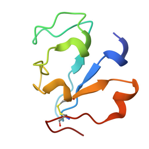 | |
UniProt | |||||
Find proteins for Q726L3 (Desulfovibrio vulgaris (strain ATCC 29579 / DSM 644 / NCIMB 8303 / VKM B-1760 / Hildenborough)) Explore Q726L3 Go to UniProtKB: Q726L3 | |||||
Entity Groups | |||||
| Sequence Clusters | 30% Identity50% Identity70% Identity90% Identity95% Identity100% Identity | ||||
| UniProt Group | Q726L3 | ||||
Sequence AnnotationsExpand | |||||
| |||||
| Ligands 2 Unique | |||||
|---|---|---|---|---|---|
| ID | Chains | Name / Formula / InChI Key | 2D Diagram | 3D Interactions | |
| PO4 (Subject of Investigation/LOI) Query on PO4 | C [auth A], E [auth B] | PHOSPHATE ION O4 P NBIIXXVUZAFLBC-UHFFFAOYSA-K |  | ||
| FE (Subject of Investigation/LOI) Query on FE | D [auth A], F [auth B] | FE (III) ION Fe VTLYFUHAOXGGBS-UHFFFAOYSA-N |  | ||
| Length ( Å ) | Angle ( ˚ ) |
|---|---|
| a = 36.565 | α = 90 |
| b = 57.856 | β = 90 |
| c = 59.198 | γ = 90 |
| Software Name | Purpose |
|---|---|
| PHENIX | refinement |
| HKL-2000 | data reduction |
| SCALA | data scaling |
| MOLREP | phasing |
| Funding Organization | Location | Grant Number |
|---|---|---|
| Not funded | -- |
RCSB PDB (citation) is hosted by
RCSB PDB is a member of the

