


Crystal structure of the 4,5-DOPA-extradiol-dioxygenase from Beta vulgaris
Chiang, C.C., Hsu, C.H.To be published.
Experimental Data Snapshot
wwPDB Validation 3D Report Full Report

Entity ID: 1 | |||||
|---|---|---|---|---|---|
| Molecule | Chains | Sequence Length | Organism | Details | Image |
| 4,5-DOPA dioxygenase extradiol | 276 | Beta vulgaris | Mutation(s): 0 Gene Names: DODA EC: 1.13.11.29 | 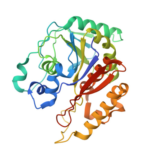 | |
UniProt | |||||
Find proteins for Q70FG7 (Beta vulgaris) Explore Q70FG7 Go to UniProtKB: Q70FG7 | |||||
Entity Groups | |||||
| Sequence Clusters | 30% Identity50% Identity70% Identity90% Identity95% Identity100% Identity | ||||
| UniProt Group | Q70FG7 | ||||
Sequence AnnotationsExpand | |||||
| |||||
| Ligands 1 Unique | |||||
|---|---|---|---|---|---|
| ID | Chains | Name / Formula / InChI Key | 2D Diagram | 3D Interactions | |
| FE (Subject of Investigation/LOI) Query on FE | C [auth A], D [auth B] | FE (III) ION Fe VTLYFUHAOXGGBS-UHFFFAOYSA-N |  | ||
| Length ( Å ) | Angle ( ˚ ) |
|---|---|
| a = 95.873 | α = 90 |
| b = 95.873 | β = 90 |
| c = 124.908 | γ = 120 |
| Software Name | Purpose |
|---|---|
| PHENIX | refinement |
| HKL-2000 | data reduction |
| HKL-2000 | data scaling |
| PHASER | phasing |
| Funding Organization | Location | Grant Number |
|---|---|---|
| Ministry of Science and Technology (MoST, Taiwan) | Taiwan | 111-2113-M-002-015-MY3 |
| Ministry of Science and Technology (MoST, Taiwan) | Taiwan | 111-2311-B-002-008-MY3 |
RCSB PDB (citation) is hosted by
RCSB PDB is a member of the

