


Crystal Structure of VibMO1
Ying, Z., Feng, K.N.To be published.
Experimental Data Snapshot
Entity ID: 1 | |||||
|---|---|---|---|---|---|
| Molecule | Chains | Sequence Length | Organism | Details | Image |
| 4-hydroxybenzoate decarboxylase | A [auth G], B [auth H], C [auth I], D [auth J] | 465 | Boreostereum vibrans | Mutation(s): 0 Gene Names: MO1 | 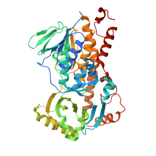 |
UniProt | |||||
Find proteins for A0A167KUL3 (Boreostereum vibrans) Explore A0A167KUL3 Go to UniProtKB: A0A167KUL3 | |||||
Entity Groups | |||||
| Sequence Clusters | 30% Identity50% Identity70% Identity90% Identity95% Identity100% Identity | ||||
| UniProt Group | A0A167KUL3 | ||||
Sequence AnnotationsExpand | |||||
| |||||
| Ligands 1 Unique | |||||
|---|---|---|---|---|---|
| ID | Chains | Name / Formula / InChI Key | 2D Diagram | 3D Interactions | |
| FAD Query on FAD | E [auth G], F [auth I] | FLAVIN-ADENINE DINUCLEOTIDE C27 H33 N9 O15 P2 VWWQXMAJTJZDQX-UYBVJOGSSA-N |  | ||
| Length ( Å ) | Angle ( ˚ ) |
|---|---|
| a = 120.43 | α = 90 |
| b = 127.78 | β = 90 |
| c = 146.45 | γ = 90 |
| Software Name | Purpose |
|---|---|
| PHENIX | refinement |
| HKL-3000 | data reduction |
| HKL-3000 | data scaling |
| PHASER | phasing |
| Funding Organization | Location | Grant Number |
|---|---|---|
| National Natural Science Foundation of China (NSFC) | China | -- |
RCSB PDB (citation) is hosted by
RCSB PDB is a member of the

