8GB3

Structure of the Mycobacterium tuberculosis Hsp70 protein DnaK bound to the nucleotide exchange factor GrpE


Experimental Data Snapshot

  • Method: ELECTRON MICROSCOPY
  • Resolution: 3.70 Å
  • Aggregation State: PARTICLE 
  • Reconstruction Method: SINGLE PARTICLE 

wwPDB Validation   3D Report Full Report


This is version 1.2 of the entry. See complete history


Literature

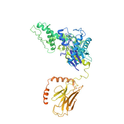
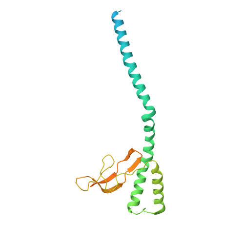
Structure of the M. tuberculosis DnaK-GrpE complex reveals how key DnaK roles are controlled.

Xiao, X.Fay, A.Molina, P.S.Kovach, A.Glickman, M.S.Li, H.

(2024) Nat Commun 15: 660-660

  • DOI: https://doi.org/10.1038/s41467-024-44933-9
  • Primary Citation of Related Structures:  
    8GB3

  • PubMed Abstract: 

    The molecular chaperone DnaK is essential for viability of Mycobacterium tuberculosis (Mtb). DnaK hydrolyzes ATP to fold substrates, and the resulting ADP is exchanged for ATP by the nucleotide exchange factor GrpE. It has been unclear how GrpE couples DnaK's nucleotide exchange with substrate release. Here we report a cryo-EM analysis of GrpE bound to an intact Mtb DnaK, revealing an asymmetric 1:2 DnaK-GrpE complex. The GrpE dimer ratchets to modulate both DnaK nucleotide-binding domain and the substrate-binding domain. We further show that the disordered GrpE N-terminus is critical for substrate release, and that the DnaK-GrpE interface is essential for protein folding activity both in vitro and in vivo. Therefore, the Mtb GrpE dimer allosterically regulates DnaK to concomitantly release ADP in the nucleotide-binding domain and substrate peptide in the substrate-binding domain.


  • Organizational Affiliation

    Department of Structural Biology, Van Andel Institute, Grand Rapids, MI, USA.


Macromolecules
Find similar proteins by:  (by identity cutoff)  |  3D Structure
Entity ID: 1
MoleculeChains Sequence LengthOrganismDetailsImage
Chaperone protein DnaKA [auth D]625Mycobacterium tuberculosisMutation(s): 0 
Gene Names: dnaK_2dnaKE5M05_19850ERS007703_00955ERS023446_02581ERS027651_01905FCN16_12395SAMEA2682864_01182SAMEA2683035_02658
UniProt
Find proteins for P9WMJ9 (Mycobacterium tuberculosis (strain ATCC 25618 / H37Rv))
Explore P9WMJ9 
Go to UniProtKB:  P9WMJ9
Entity Groups  
Sequence Clusters30% Identity50% Identity70% Identity90% Identity95% Identity100% Identity
UniProt GroupP9WMJ9
Sequence Annotations
Expand
  • Reference Sequence
Find similar proteins by:  (by identity cutoff)  |  3D Structure
Entity ID: 2
MoleculeChains Sequence LengthOrganismDetailsImage
Protein GrpEB [auth E],
C [auth F]
235Mycobacterium tuberculosisMutation(s): 0 
Gene Names: grpEE5M05_19845E5M52_19255E5M78_19290ERS007681_03281ERS007703_00954ERS024276_02384SAMEA2683035_02659
UniProt
Find proteins for P9WMT5 (Mycobacterium tuberculosis (strain ATCC 25618 / H37Rv))
Explore P9WMT5 
Go to UniProtKB:  P9WMT5
Entity Groups  
Sequence Clusters30% Identity50% Identity70% Identity90% Identity95% Identity100% Identity
UniProt GroupP9WMT5
Sequence Annotations
Expand
  • Reference Sequence
Experimental Data & Validation

Experimental Data

  • Method: ELECTRON MICROSCOPY
  • Resolution: 3.70 Å
  • Aggregation State: PARTICLE 
  • Reconstruction Method: SINGLE PARTICLE 
EM Software:
TaskSoftware PackageVersion
RECONSTRUCTIONcryoSPARC
MODEL REFINEMENTPHENIX

Structure Validation

View Full Validation Report


Entry History & Funding Information

Deposition Data


Funding OrganizationLocationGrant Number
National Institutes of Health/National Library of Medicine (NIH/NLM)United StatesR01

Revision History  (Full details and data files)

  • Version 1.0: 2024-01-31
    Type: Initial release
  • Version 1.1: 2024-03-13
    Changes: Database references
  • Version 1.2: 2024-05-01
    Changes: Database references