Conformational dynamics control assembly of an extremely long bacteriophage tail tube.
Agnello, E., Pajak, J., Liu, X., Kelch, B.A.(2023) J Biol Chem 299: 103021-103021
- PubMed: 36791911
- DOI: https://doi.org/10.1016/j.jbc.2023.103021
- Primary Citation of Related Structures:
8ED0, 8EDX - PubMed Abstract:
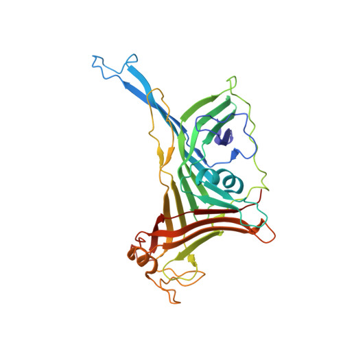
Tail tube assembly is an essential step in the lifecycle of long-tailed bacteriophages. Limited structural and biophysical information has impeded an understanding of assembly and stability of their long, flexible tail tubes. The hyperthermophilic phage P74-26 is particularly intriguing as it has the longest tail of any known virus (nearly 1 μm) and is the most thermostable known phage. Here, we use structures of the P74-26 tail tube along with an in vitro system for studying tube assembly kinetics to propose the first molecular model for the tail tube assembly of long-tailed phages. Our high-resolution cryo-EM structure provides insight into how the P74-26 phage assembles through flexible loops that fit into neighboring rings through tight "ball-and-socket"-like interactions. Guided by this structure, and in combination with mutational, light scattering, and molecular dynamics simulations data, we propose a model for the assembly of conserved tube-like structures across phage and other entities possessing tail tube-like proteins. We propose that formation of a full ring promotes the adoption of a tube elongation-competent conformation among the flexible loops and their corresponding sockets, which is further stabilized by an adjacent ring. Tail assembly is controlled by the cooperative interaction of dynamic intraring and interring contacts. Given the structural conservation among tail tube proteins and tail-like structures, our model can explain the mechanism of high-fidelity assembly of long, stable tubes.
Organizational Affiliation:
Department of Biochemistry and Molecular Biotechnology, University of Massachusetts Chan Medical School, Worcester Massachusetts, USA.