8E62

STRUCTURE OF Pcryo_0615 from Psychrobacter cryohalolentis, an N-acetyltransferase required to produce Diacetamido-2,3-dideoxy-D-glucuronic acid


Experimental Data Snapshot

  • Method: X-RAY DIFFRACTION
  • Resolution: 1.80 Å
  • R-Value Free: 0.208 
  • R-Value Work: 0.166 
  • R-Value Observed: 0.168 

wwPDB Validation   3D Report Full Report


Ligand Structure Quality Assessment 


This is version 1.2 of the entry. See complete history


Literature

Investigation of the enzymes required for the biosynthesis of 2,3-diacetamido-2,3-dideoxy-d-glucuronic acid in Psychrobacter cryohalolentis K5 T.

Hofmeister, D.L.Seltzner, C.A.Bockhaus, N.J.Thoden, J.B.Holden, H.M.

(2023) Protein Sci 32: e4502-e4502

  • DOI: https://doi.org/10.1002/pro.4502
  • Primary Citation of Related Structures:  
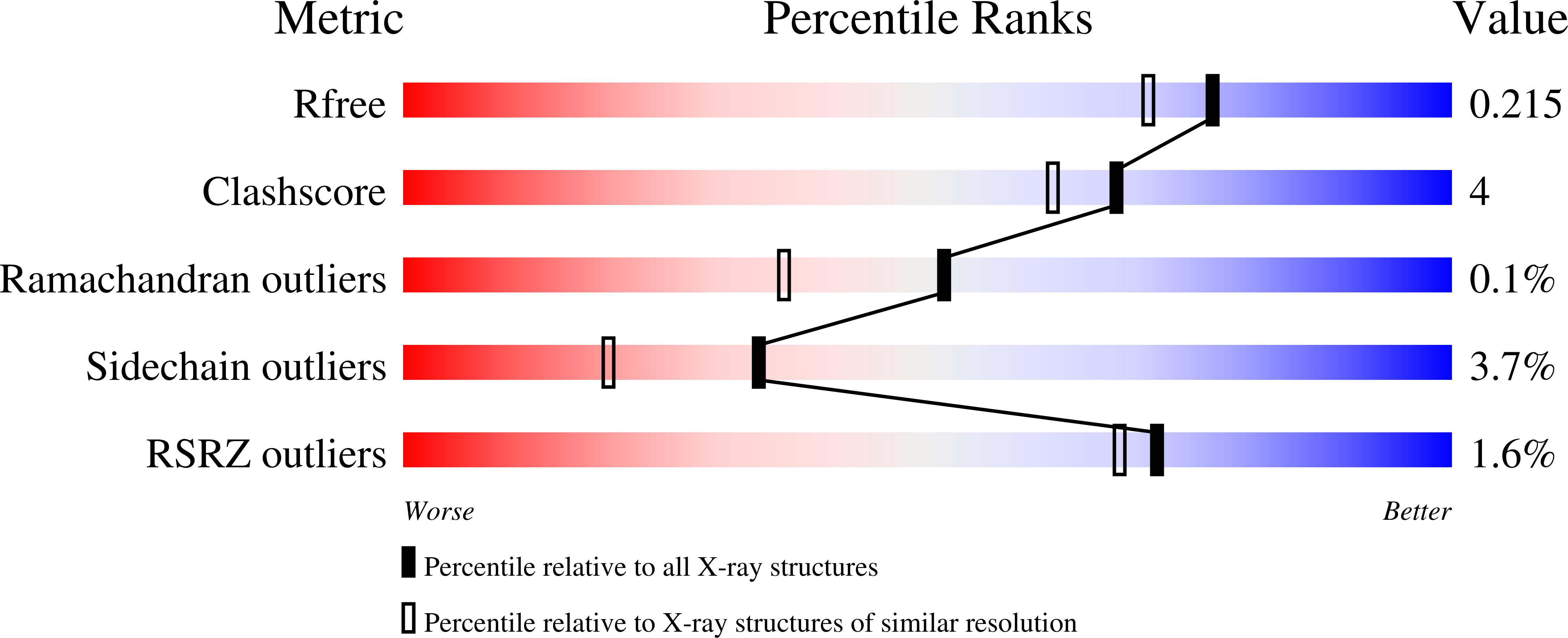
    8E62, 8E75, 8E77

  • PubMed Abstract: 

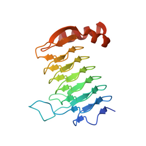
    Psychrobacter cryohalolentis K5 T is a Gram-negative bacterium first isolated from Siberian permafrost in 2006. It has a complex O-antigen containing l-rhamnose, d-galactose, two diacetamido-sugars, and one triacetamido-sugar. The biosynthetic pathway for one of the diacetamido-sugars, namely 2,3-diacetamido-2,3-dideoxy-d-glucuronic acid, is presently unknown. Utilizing the published genome sequence of P. cryohalolentis K5 T , we hypothesized that the genes designated Pcryo_0613, Pcryo_0614, Pcryo_0616, and Pcryo_0615 encode for a uridine dinucleotide (UDP)-N-acetyl-d-glucosamine 6-dehydrogenase, an nicotinamide adenine dinucleotide (oxidized) (NAD + )-dependent dehydrogenase, a pyridoxal 5'-phosphate (PLP)-dependent aminotransferase, and an N-acetyltransferase, respectively, activities of which would be required for the biosynthesis of this unusual carbohydrate. Here we present the cloning, overexpression, and purification of these hypothetical proteins. Kinetic data on the enzymes encoded by Pcryo_0613, Pcryo_0614, and Pcryo_0615 confirmed their postulated biochemical activities. In addition, the high-resolution X-ray structures of both the internal and external aldimine forms of the aminotransferase were determined to 1.25 and 1.0 Å, respectively. Finally, the three-dimensional architecture of the N-acetyltransferase in complex with its substrate and coenzyme A was solved to 1.8 Å resolution. Strikingly, the N-acetyltransferase was shown to adopt a new motif for UDP-sugar binding. The data presented herein provide additional insight into sugar biosynthesis in Gram-negative bacteria.


  • Organizational Affiliation

    Department of Biochemistry, University of Wisconsin, Madison, Wisconsin, USA.


Macromolecules
Find similar proteins by:  (by identity cutoff)  |  3D Structure
Entity ID: 1
MoleculeChains Sequence LengthOrganismDetailsImage
UDP-N-acetylglucosamine acyltransferase
A, B, C, D, E
A, B, C, D, E, F
189Psychrobacter cryohalolentis K5Mutation(s): 0 
Gene Names: Pcryo_0615
UniProt
Find proteins for Q1QD55 (Psychrobacter cryohalolentis (strain ATCC BAA-1226 / DSM 17306 / VKM B-2378 / K5))
Explore Q1QD55 
Go to UniProtKB:  Q1QD55
Entity Groups  
Sequence Clusters30% Identity50% Identity70% Identity90% Identity95% Identity100% Identity
UniProt GroupQ1QD55
Sequence Annotations
Expand
  • Reference Sequence
Small Molecules
Ligands 3 Unique
IDChains Name / Formula / InChI Key2D Diagram3D Interactions
COA (Subject of Investigation/LOI)
Query on COA

Download Ideal Coordinates CCD File 
H [auth A]
I [auth A]
K [auth B]
N [auth E]
O [auth E]
H [auth A],
I [auth A],
K [auth B],
N [auth E],
O [auth E],
P [auth F]
COENZYME A
C21 H36 N7 O16 P3 S
RGJOEKWQDUBAIZ-IBOSZNHHSA-N
MJZ (Subject of Investigation/LOI)
Query on MJZ

Download Ideal Coordinates CCD File 
G [auth A],
J [auth B],
Q [auth F]
(2S,3S,4R,5R,6R)-5-(acetylamino)-4-amino-6-{[(R)-{[(R)-{[(2R,3S,4R,5R)-5-(2,4-dioxo-3,4-dihydropyrimidin-1(2H)-yl)-3,4-dihydroxytetrahydrofuran-2-yl]methoxy}(hydroxy)phosphoryl]oxy}(hydroxy)phosphoryl]oxy}-3-hydroxytetrahydro-2H-pyran-2-carboxylic acid
C17 H26 N4 O17 P2
RRAQYLXLCYIZBB-HHKCBAECSA-N
NA
Query on NA

Download Ideal Coordinates CCD File 
L [auth B],
M [auth D]
SODIUM ION
Na
FKNQFGJONOIPTF-UHFFFAOYSA-N
Experimental Data & Validation

Experimental Data

  • Method: X-RAY DIFFRACTION
  • Resolution: 1.80 Å
  • R-Value Free: 0.208 
  • R-Value Work: 0.166 
  • R-Value Observed: 0.168 
  • Space Group: P 1 21 1
Unit Cell:
Length ( Å )Angle ( ˚ )
a = 82.113α = 90
b = 79.768β = 114.28
c = 96.616γ = 90
Software Package:
Software NamePurpose
REFMACrefinement
PDB_EXTRACTdata extraction
HKL-3000data reduction
HKL-3000data scaling
PHASERphasing

Structure Validation

View Full Validation Report


Ligand Structure Quality Assessment 


Entry History & Funding Information

Deposition Data


Funding OrganizationLocationGrant Number
National Institutes of Health/National Institute of General Medical Sciences (NIH/NIGMS)United StatesR35 GM134643

Revision History  (Full details and data files)

  • Version 1.0: 2022-11-23
    Type: Initial release
  • Version 1.1: 2023-01-11
    Changes: Database references
  • Version 1.2: 2023-10-25
    Changes: Data collection, Refinement description