8DPB

MeaB in complex with the cobalamin-binding domain of its target mutase with GMPPCP bound


Experimental Data Snapshot

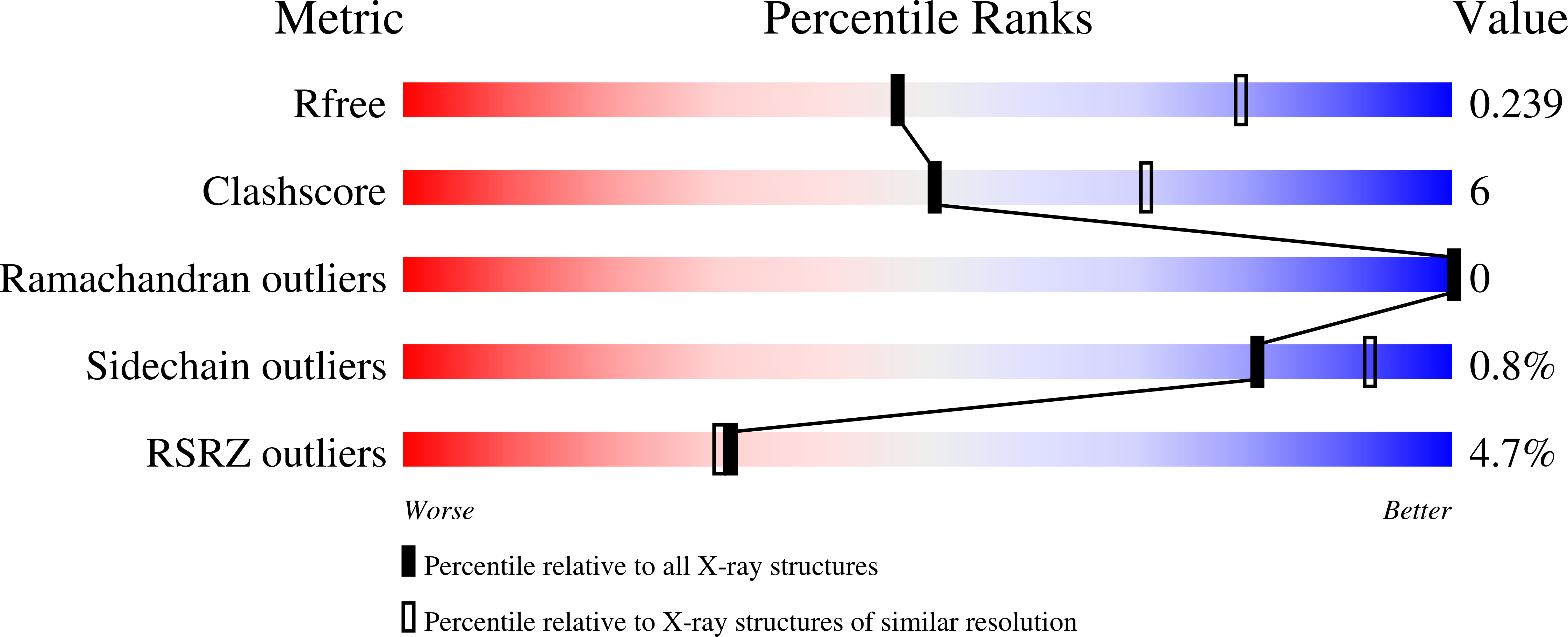
  • Method: X-RAY DIFFRACTION
  • Resolution: 2.72 Å
  • R-Value Free: 0.237 
  • R-Value Work: 0.216 
  • R-Value Observed: 0.217 

wwPDB Validation   3D Report Full Report


Ligand Structure Quality Assessment 


This is version 1.1 of the entry. See complete history


Literature

Structure of metallochaperone in complex with the cobalamin-binding domain of its target mutase provides insight into cofactor delivery.

Vaccaro, F.A.Born, D.A.Drennan, C.L.

(2023) Proc Natl Acad Sci U S A 120: e2214085120-e2214085120

  • DOI: https://doi.org/10.1073/pnas.2214085120
  • Primary Citation of Related Structures:  
    8DPB

  • PubMed Abstract: 

    G-protein metallochaperone MeaB in bacteria [methylmalonic aciduria type A (MMAA) in humans] is responsible for facilitating the delivery of adenosylcobalamin (AdoCbl) to methylmalonyl-CoA mutase (MCM), the only AdoCbl-dependent enzyme in humans. Genetic defects in the switch III region of MMAA lead to the genetic disorder methylmalonic aciduria in which the body is unable to process certain lipids. Here, we present a crystal structure of Methylobacterium extorquens MeaB bound to a nonhydrolyzable guanosine triphosphate (GTP) analog guanosine-5'-[(β,γ)-methyleno]triphosphate (GMPPCP) with the Cbl-binding domain of its target mutase enzyme ( Me MCM cbl ). This structure provides an explanation for the stimulation of the GTP hydrolyase activity of MeaB afforded by target protein binding. We find that upon MCM cbl association, one protomer of the MeaB dimer rotates ~180°, such that the inactive state of MeaB is converted to an active state in which the nucleotide substrate is now surrounded by catalytic residues. Importantly, it is the switch III region that undergoes the largest change, rearranging to make direct contacts with the terminal phosphate of GMPPCP. These structural data additionally provide insights into the molecular basis by which this metallochaperone contributes to AdoCbl delivery without directly binding the cofactor. Our data suggest a model in which GTP-bound MeaB stabilizes a conformation of MCM that is open for AdoCbl insertion, and GTP hydrolysis, as signaled by switch III residues, allows MCM to close and trap its cofactor. Substitutions of switch III residues destabilize the active state of MeaB through loss of protein:nucleotide and protein:protein interactions at the dimer interface, thus uncoupling GTP hydrolysis from AdoCbl delivery.


  • Organizational Affiliation

    Department of Chemistry, Massachusetts Institute of Technology, Cambridge, MA 01239.


Macromolecules
Find similar proteins by:  (by identity cutoff)  |  3D Structure
Entity ID: 1
MoleculeChains Sequence LengthOrganismDetailsImage
Methylmalonyl-CoA mutase accessory protein
A, B
329Methylorubrum extorquens AM1Mutation(s): 0 
Gene Names: meaBMexAM1_META1p0188
UniProt
Find proteins for C5AP93 (Methylorubrum extorquens (strain ATCC 14718 / DSM 1338 / JCM 2805 / NCIMB 9133 / AM1))
Explore C5AP93 
Go to UniProtKB:  C5AP93
Entity Groups  
Sequence Clusters30% Identity50% Identity70% Identity90% Identity95% Identity100% Identity
UniProt GroupC5AP93
Sequence Annotations
Expand
  • Reference Sequence
Find similar proteins by:  (by identity cutoff)  |  3D Structure
Entity ID: 2
MoleculeChains Sequence LengthOrganismDetailsImage
Methylmalonyl-CoA mutase, alpha subunit
C, D
191Methylorubrum extorquens AM1Mutation(s): 0 
Gene Names: mcmAMexAM1_META1p5251
EC: 5.4.99.2
UniProt
Find proteins for C5AV67 (Methylorubrum extorquens (strain ATCC 14718 / DSM 1338 / JCM 2805 / NCIMB 9133 / AM1))
Explore C5AV67 
Go to UniProtKB:  C5AV67
Entity Groups  
Sequence Clusters30% Identity50% Identity70% Identity90% Identity95% Identity100% Identity
UniProt GroupC5AV67
Sequence Annotations
Expand
  • Reference Sequence
Experimental Data & Validation

Experimental Data

  • Method: X-RAY DIFFRACTION
  • Resolution: 2.72 Å
  • R-Value Free: 0.237 
  • R-Value Work: 0.216 
  • R-Value Observed: 0.217 
  • Space Group: P 21 21 21
Unit Cell:
Length ( Å )Angle ( ˚ )
a = 66.23α = 90
b = 80.98β = 90
c = 166.38γ = 90
Software Package:
Software NamePurpose
PHASERphasing
XDSdata reduction
XDSdata scaling
PHENIXrefinement

Structure Validation

View Full Validation Report


Ligand Structure Quality Assessment 


Entry History & Funding Information

Deposition Data


Funding OrganizationLocationGrant Number
National Institutes of Health/National Institute of General Medical Sciences (NIH/NIGMS)United StatesR35 GM126982
Howard Hughes Medical Institute (HHMI)United States--
National Institutes of Health/National Institute of General Medical Sciences (NIH/NIGMS)United StatesF31 GM131648
National Institutes of Health/National Institute of General Medical Sciences (NIH/NIGMS)United StatesT32 GM008313

Revision History  (Full details and data files)

  • Version 1.0: 2023-03-01
    Type: Initial release
  • Version 1.1: 2024-04-03
    Changes: Data collection, Refinement description