8BNX

Crystal structure of Pif1 from Deferribacter desulfuricans in complex with AMPPNP


Experimental Data Snapshot

  • Method: X-RAY DIFFRACTION
  • Resolution: 3.12 Å
  • R-Value Free: 0.255 
  • R-Value Work: 0.240 
  • R-Value Observed: 0.241 

wwPDB Validation   3D Report Full Report


Ligand Structure Quality Assessment 


This is version 1.1 of the entry. See complete history


Literature

Structural Studies of Pif1 Helicases from Thermophilic Bacteria.

Rety, S.Zhang, Y.Fu, W.Wang, S.Chen, W.F.Xi, X.G.

(2023) Microorganisms 11

  • DOI: https://doi.org/10.3390/microorganisms11020479
  • Primary Citation of Related Structures:  
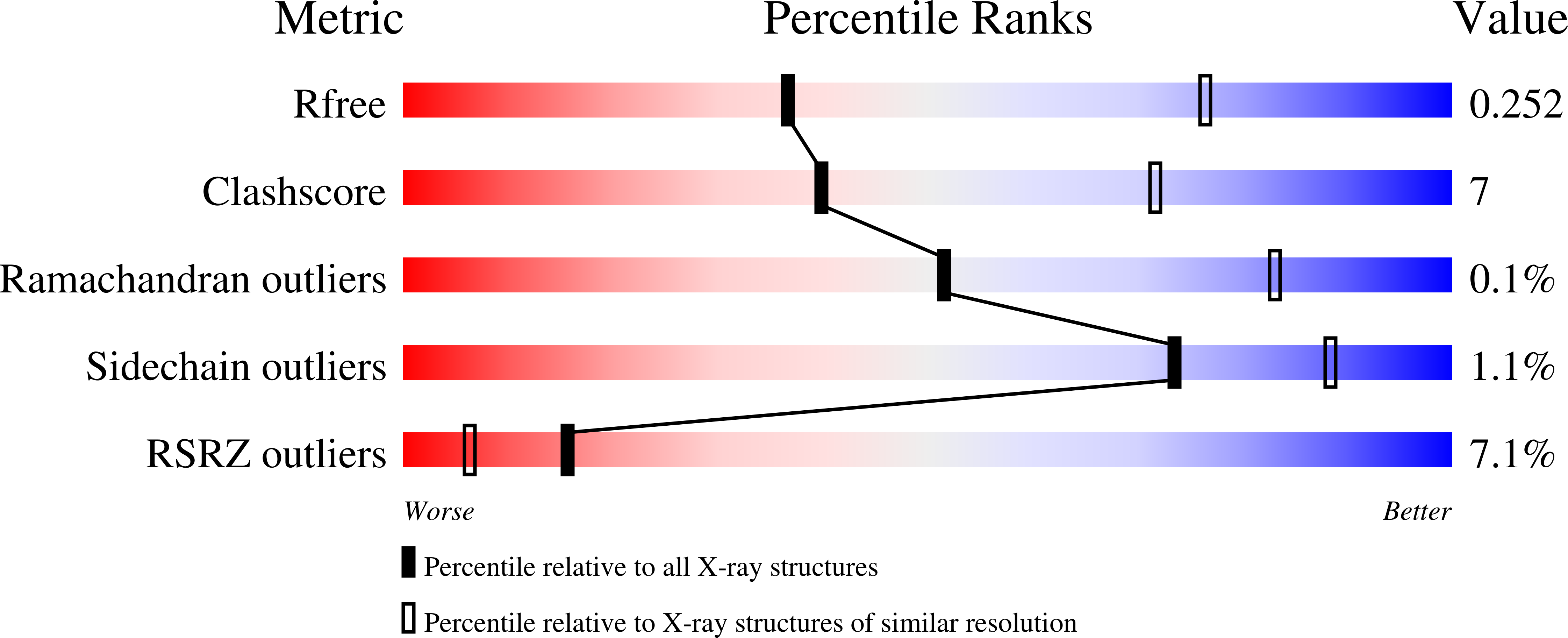
    8BNS, 8BNV, 8BNX

  • PubMed Abstract: 

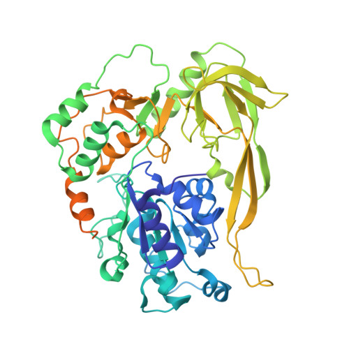
    Pif1 proteins are DNA helicases belonging to Superfamily 1, with 5' to 3' directionality. They are conserved from bacteria to human and have been shown to be particularly important in eukaryotes for replication and nuclear and mitochondrial genome stability. However, Pif1 functions in bacteria are less known. While most Pif1 from mesophilic bacteria consist of the helicase core with limited N-terminal and C-terminal extensions, some Pif1 from thermophilic bacteria exhibit a C-terminal WYL domain. We solved the crystal structures of Pif1 helicase cores from thermophilic bacteria Deferribacter desulfuricans and Sulfurihydrogenibium sp. in apo and nucleotide bound form. We show that the N-terminal part is important for ligand binding. The full-length Pif1 helicase was predicted based on the Alphafold algorithm and the nucleic acid binding on the Pif1 helicase core and the WYL domain was modelled based on known crystallographic structures. The model predicts that amino acids in the domains 1A, WYL, and linker between the Helicase core and WYL are important for nucleic acid binding. Therefore, the N-terminal and C-terminal extensions may be necessary to strengthen the binding of nucleic acid on these Pif1 helicases. This may be an adaptation to thermophilic conditions.


  • Organizational Affiliation

    Laboratoire de Biologie et Modelisation de la Cellule, Ecole Normale Superieure de Lyon, CNRS, UMR 5239, Inserm, U1293, Universite Claude Bernard Lyon 1, 46 allee d'Italie, F-69364 Lyon, France.


Macromolecules
Find similar proteins by:  (by identity cutoff)  |  3D Structure
Entity ID: 1
MoleculeChains Sequence LengthOrganismDetailsImage
AAA family ATPase
A, B
510Deferribacter desulfuricansMutation(s): 0 
Gene Names: DEFDS_0256
UniProt
Find proteins for D3PAZ1 (Deferribacter desulfuricans (strain DSM 14783 / JCM 11476 / NBRC 101012 / SSM1))
Explore D3PAZ1 
Go to UniProtKB:  D3PAZ1
Entity Groups  
Sequence Clusters30% Identity50% Identity70% Identity90% Identity95% Identity100% Identity
UniProt GroupD3PAZ1
Sequence Annotations
Expand
  • Reference Sequence
Small Molecules
Ligands 1 Unique
IDChains Name / Formula / InChI Key2D Diagram3D Interactions
ANP (Subject of Investigation/LOI)
Query on ANP

Download Ideal Coordinates CCD File 
C [auth A]PHOSPHOAMINOPHOSPHONIC ACID-ADENYLATE ESTER
C10 H17 N6 O12 P3
PVKSNHVPLWYQGJ-KQYNXXCUSA-N
Experimental Data & Validation

Experimental Data

  • Method: X-RAY DIFFRACTION
  • Resolution: 3.12 Å
  • R-Value Free: 0.255 
  • R-Value Work: 0.240 
  • R-Value Observed: 0.241 
  • Space Group: P 21 21 2
Unit Cell:
Length ( Å )Angle ( ˚ )
a = 143.797α = 90
b = 151.376β = 90
c = 51.857γ = 90
Software Package:
Software NamePurpose
PHENIXrefinement
PHENIXrefinement
autoPROCdata reduction
autoPROCdata scaling
PHASERphasing

Structure Validation

View Full Validation Report


Ligand Structure Quality Assessment 


Entry History & Funding Information

Deposition Data


Funding OrganizationLocationGrant Number
Centre National de la Recherche Scientifique (CNRS)France--

Revision History  (Full details and data files)

  • Version 1.0: 2023-03-08
    Type: Initial release
  • Version 1.1: 2024-02-07
    Changes: Data collection, Refinement description