8AJY

Ruminococcus flavefaciens Cohesin-Dockerin structure: dockerin from ScaH adaptor scaffoldin in complex with the cohesin from ScaE anchoring scaffoldin


Experimental Data Snapshot

  • Method: X-RAY DIFFRACTION
  • Resolution: 1.71 Å
  • R-Value Free: 0.237 
  • R-Value Work: 0.194 
  • R-Value Observed: 0.196 

wwPDB Validation   3D Report Full Report


This is version 1.2 of the entry. See complete history


Literature

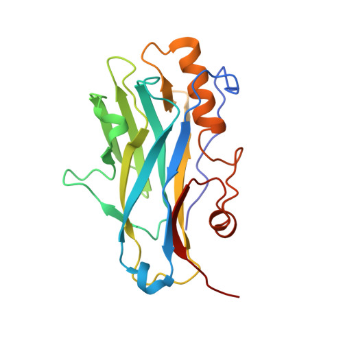
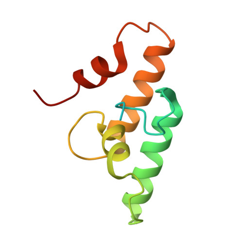
Structure-function studies can improve binding affinity of cohesin-dockerin interactions for multi-protein assemblies.

Duarte, M.Alves, V.D.Correia, M.Caseiro, C.Ferreira, L.M.A.Romao, M.J.Carvalho, A.L.Najmudin, S.Bayer, E.A.Fontes, C.M.G.A.Bule, P.

(2023) Int J Biol Macromol 224: 55-67

  • DOI: https://doi.org/10.1016/j.ijbiomac.2022.10.102
  • Primary Citation of Related Structures:  
    8AJY

  • PubMed Abstract: 

    The cellulosome is an elaborate multi-enzyme structure secreted by many anaerobic microorganisms for the efficient degradation of lignocellulosic substrates. It is composed of multiple catalytic and non-catalytic components that are assembled through high-affinity protein-protein interactions between the enzyme-borne dockerin (Doc) modules and the repeated cohesin (Coh) modules present in primary scaffoldins. In some cellulosomes, primary scaffoldins can interact with adaptor and cell-anchoring scaffoldins to create structures of increasing complexity. The cellulosomal system of the ruminal bacterium, Ruminococcus flavefaciens, is one of the most intricate described to date. An unprecedent number of different Doc specificities results in an elaborate architecture, assembled exclusively through single-binding-mode type-III Coh-Doc interactions. However, a set of type-III Docs exhibits certain features associated with the classic dual-binding mode Coh-Doc interaction. Here, the structure of the adaptor scaffoldin-borne ScaH Doc in complex with the Coh from anchoring scaffoldin ScaE is described. This complex, unlike previously described type-III interactions in R. flavefaciens, was found to interact in a dual-binding mode. The key residues determining Coh recognition were also identified. This information was used to perform structure-informed protein engineering to change the electrostatic profile of the binding surface and to improve the affinity between the two modules. The results show that the nature of the residues in the ligand-binding surface plays a major role in Coh recognition and that Coh-Doc affinity can be manipulated through rational design, a key feature for the creation of designer cellulosomes or other affinity-based technologies using tailored Coh-Doc interactions.


  • Organizational Affiliation

    CIISA-Centre for Interdisciplinary Research in Animal Health, Faculty of Veterinary Medicine, University of Lisbon, 1300-477 Lisbon, Portugal; Associate Laboratory for Animal and Veterinary Sciences (AL4AnimalS), 1300-477 Lisbon, Portugal.


Macromolecules
Find similar proteins by:  (by identity cutoff)  |  3D Structure
Entity ID: 1
MoleculeChains Sequence LengthOrganismDetailsImage
Cell-wall anchoring protein
A, C
203Ruminococcus flavefaciens FD-1Mutation(s): 0 
Gene Names: scaE
UniProt
Find proteins for A0AEF6 (Ruminococcus flavefaciens)
Explore A0AEF6 
Go to UniProtKB:  A0AEF6
Entity Groups  
Sequence Clusters30% Identity50% Identity70% Identity90% Identity95% Identity100% Identity
UniProt GroupA0AEF6
Sequence Annotations
Expand
  • Reference Sequence
Find similar proteins by:  (by identity cutoff)  |  3D Structure
Entity ID: 2
MoleculeChains Sequence LengthOrganismDetailsImage
Dockerin from ScaH
B, D
112Ruminococcus flavefaciens FD-1Mutation(s): 0 
Entity Groups  
Sequence Clusters30% Identity50% Identity70% Identity90% Identity95% Identity100% Identity
Sequence Annotations
Expand
  • Reference Sequence
Small Molecules
Ligands 3 Unique
IDChains Name / Formula / InChI Key2D Diagram3D Interactions
SO4
Query on SO4

Download Ideal Coordinates CCD File 
G [auth A]
H [auth A]
I [auth A]
J [auth A]
K [auth A]
G [auth A],
H [auth A],
I [auth A],
J [auth A],
K [auth A],
L [auth A],
O [auth B],
P [auth B],
S [auth C],
T [auth C],
U [auth C],
V [auth C],
Y [auth D]
SULFATE ION
O4 S
QAOWNCQODCNURD-UHFFFAOYSA-L
GOL
Query on GOL

Download Ideal Coordinates CCD File 
F [auth A],
R [auth C]
GLYCEROL
C3 H8 O3
PEDCQBHIVMGVHV-UHFFFAOYSA-N
CA (Subject of Investigation/LOI)
Query on CA

Download Ideal Coordinates CCD File 
E [auth A]
M [auth B]
N [auth B]
Q [auth C]
W [auth D]
E [auth A],
M [auth B],
N [auth B],
Q [auth C],
W [auth D],
X [auth D]
CALCIUM ION
Ca
BHPQYMZQTOCNFJ-UHFFFAOYSA-N
Experimental Data & Validation

Experimental Data

  • Method: X-RAY DIFFRACTION
  • Resolution: 1.71 Å
  • R-Value Free: 0.237 
  • R-Value Work: 0.194 
  • R-Value Observed: 0.196 
  • Space Group: P 1 21 1
Unit Cell:
Length ( Å )Angle ( ˚ )
a = 40.81α = 90
b = 120β = 97.77
c = 65.95γ = 90
Software Package:
Software NamePurpose
REFMACrefinement
PDB_EXTRACTdata extraction
autoPROCdata reduction
autoPROCdata scaling
PHASERphasing

Structure Validation

View Full Validation Report


Entry History & Funding Information

Deposition Data


Funding OrganizationLocationGrant Number
Foundation for Science and Technology (FCT)United Kingdom--

Revision History  (Full details and data files)

  • Version 1.0: 2022-11-02
    Type: Initial release
  • Version 1.1: 2022-12-28
    Changes: Database references
  • Version 1.2: 2024-01-31
    Changes: Data collection, Refinement description