


crystal structure of UBE2O
Fu, Z., Zhu, W., Huang, H.To be published.
Experimental Data Snapshot
wwPDB Validation 3D Report Full Report

Entity ID: 1 | |||||
|---|---|---|---|---|---|
| Molecule | Chains | Sequence Length | Organism | Details | Image |
| (E3-independent) E2 ubiquitin-conjugating enzyme UBE2O | 928 | Trametes pubescens | Mutation(s): 0 Gene Names: TRAPUB_10883 | 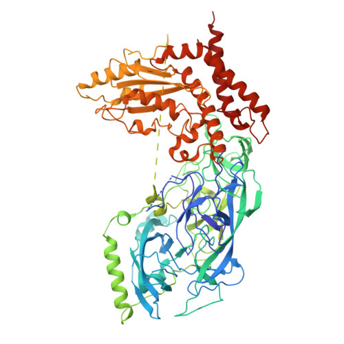 | |
UniProt | |||||
Find proteins for A0A1M2VY70 (Trametes pubescens) Explore A0A1M2VY70 Go to UniProtKB: A0A1M2VY70 | |||||
Entity Groups | |||||
| Sequence Clusters | 30% Identity50% Identity70% Identity90% Identity95% Identity100% Identity | ||||
| UniProt Group | A0A1M2VY70 | ||||
Sequence AnnotationsExpand | |||||
| |||||
| Length ( Å ) | Angle ( ˚ ) |
|---|---|
| a = 115.32 | α = 90 |
| b = 132.529 | β = 90 |
| c = 155.036 | γ = 90 |
| Software Name | Purpose |
|---|---|
| PHENIX | refinement |
| PHENIX | phasing |
| STARANISO | data scaling |
| PDB_EXTRACT | data extraction |
| PROCOR | data reduction |
| Funding Organization | Location | Grant Number |
|---|---|---|
| Not funded | -- |
RCSB PDB (citation) is hosted by
RCSB PDB is a member of the

