7XTJ

Crystal structure of E88A mutant of GH3 beta-xylosidase from Aspergillus niger (AnBX)


Experimental Data Snapshot

  • Method: X-RAY DIFFRACTION
  • Resolution: 2.50 Å
  • R-Value Free: 0.256 
  • R-Value Work: 0.214 
  • R-Value Observed: 0.216 

wwPDB Validation   3D Report Full Report


Ligand Structure Quality Assessment 


This is version 1.2 of the entry. See complete history


Literature

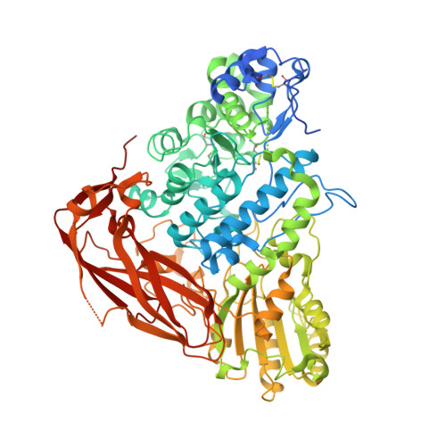
Crystal structure and identification of amino acid residues for catalysis and binding of GH3 AnBX beta-xylosidase from Aspergillus niger.

Kaenying, W.Choengpanya, K.Tagami, T.Wattana-Amorn, P.Lang, W.Okuyama, M.Li, Y.K.Kimura, A.Kongsaeree, P.T.

(2023) Appl Microbiol Biotechnol 107: 2335-2349

  • DOI: https://doi.org/10.1007/s00253-023-12445-z
  • Primary Citation of Related Structures:  
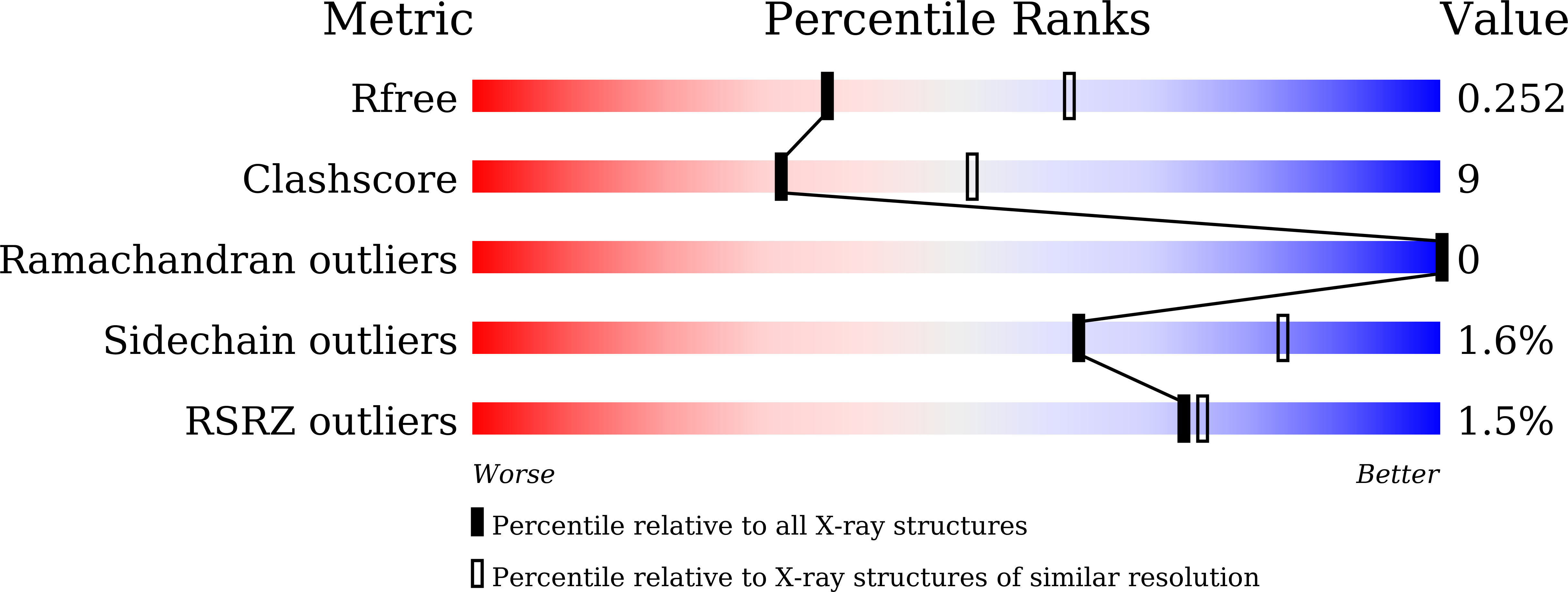
    7XTJ

  • PubMed Abstract: 

    β-Xylosidases catalyze the hydrolysis of xylooligosaccharides to xylose in the final step of hemicellulose degradation. AnBX, which is a GH3 β-xylosidase from Aspergillus niger, has a high catalytic efficiency toward xyloside substrates. In this study, we report the three-dimensional structure and the identification of catalytic and substrate binding residues of AnBX by performing site-directed mutagenesis, kinetic analysis, and NMR spectroscopy-associated analysis of the azide rescue reaction. The structure of the E88A mutant of AnBX, determined at 2.5-Å resolution, contains two molecules in the asymmetric unit, each of which is composed of three domains, namely an N-terminal (β/α) 8 TIM-barrel-like domain, an (α/β) 6 sandwich domain, and a C-terminal fibronectin type III domain. Asp288 and Glu500 of AnBX were experimentally confirmed to act as the catalytic nucleophile and acid/base catalyst, respectively. The crystal structure revealed that Trp86, Glu88 and Cys289, which formed a disulfide bond with Cys321, were located at subsite -1. Although the E88D and C289W mutations reduced catalytic efficiency toward all four substrates tested, the substitution of Trp86 with Ala, Asp and Ser increased the substrate preference for glucoside relative to xyloside substrates, indicating that Trp86 is responsible for the xyloside specificity of AnBX. The structural and biochemical information of AnBX obtained in this study provides invaluable insight into modulating the enzymatic properties for the hydrolysis of lignocellulosic biomass. KEY POINTS: • Asp288 and Glu500 of AnBX are the nucleophile and acid/base catalyst, respectively • Glu88 and the Cys289-Cys321 disulfide bond are crucial for the catalytic activity of AnBX • The W86A and W86S mutations in AnBX increased the preference for glucoside substrates.


  • Organizational Affiliation

    Department of Biochemistry, Faculty of Science, Kasetsart University, Bangkok, 10900, Thailand.


Macromolecules
Find similar proteins by:  (by identity cutoff)  |  3D Structure
Entity ID: 1
MoleculeChains Sequence LengthOrganismDetailsImage
Xylan 1,4-beta-xylosidase
A, B
788Aspergillus nigerMutation(s): 1 
EC: 3.2.1.37
UniProt
Find proteins for A0A023J5W7 (Aspergillus niger)
Explore A0A023J5W7 
Go to UniProtKB:  A0A023J5W7
Entity Groups  
Sequence Clusters30% Identity50% Identity70% Identity90% Identity95% Identity100% Identity
UniProt GroupA0A023J5W7
Sequence Annotations
Expand
  • Reference Sequence
Oligosaccharides

Help

Entity ID: 2
MoleculeChains Length2D Diagram Glycosylation3D Interactions
alpha-D-mannopyranose-(1-2)-alpha-D-mannopyranose-(1-3)-[alpha-D-mannopyranose-(1-6)]beta-D-mannopyranose-(1-4)-2-acetamido-2-deoxy-beta-D-glucopyranose-(1-4)-2-acetamido-2-deoxy-beta-D-glucopyranose
C, E
6N-Glycosylation
Glycosylation Resources
GlyTouCan:  G56014GC
GlyCosmos:  G56014GC
GlyGen:  G56014GC
Entity ID: 3
MoleculeChains Length2D Diagram Glycosylation3D Interactions
2-acetamido-2-deoxy-beta-D-glucopyranose-(1-4)-2-acetamido-2-deoxy-beta-D-glucopyranose
D
2N-Glycosylation
Glycosylation Resources
GlyTouCan:  G42666HT
GlyCosmos:  G42666HT
GlyGen:  G42666HT
Entity ID: 4
MoleculeChains Length2D Diagram Glycosylation3D Interactions
beta-D-mannopyranose-(1-4)-2-acetamido-2-deoxy-beta-D-glucopyranose-(1-4)-2-acetamido-2-deoxy-beta-D-glucopyranose
F
3N-Glycosylation
Glycosylation Resources
GlyTouCan:  G15407YE
GlyCosmos:  G15407YE
GlyGen:  G15407YE
Small Molecules
Ligands 2 Unique
IDChains Name / Formula / InChI Key2D Diagram3D Interactions
NAG
Query on NAG

Download Ideal Coordinates CCD File 
G [auth A]
H [auth A]
I [auth A]
J [auth A]
L [auth B]
G [auth A],
H [auth A],
I [auth A],
J [auth A],
L [auth B],
M [auth B],
N [auth B],
O [auth B],
P [auth B]
2-acetamido-2-deoxy-beta-D-glucopyranose
C8 H15 N O6
OVRNDRQMDRJTHS-FMDGEEDCSA-N
GOL
Query on GOL

Download Ideal Coordinates CCD File 
K [auth A],
Q [auth B]
GLYCEROL
C3 H8 O3
PEDCQBHIVMGVHV-UHFFFAOYSA-N
Experimental Data & Validation

Experimental Data

  • Method: X-RAY DIFFRACTION
  • Resolution: 2.50 Å
  • R-Value Free: 0.256 
  • R-Value Work: 0.214 
  • R-Value Observed: 0.216 
  • Space Group: P 41 21 2
Unit Cell:
Length ( Å )Angle ( ˚ )
a = 120.881α = 90
b = 120.881β = 90
c = 266.443γ = 90
Software Package:
Software NamePurpose
PHENIXrefinement
XDSdata reduction
XDSdata scaling
PHENIXphasing

Structure Validation

View Full Validation Report


Ligand Structure Quality Assessment 


Entry History & Funding Information

Deposition Data


Funding OrganizationLocationGrant Number
Other governmentThailandNRCT5-RSA63002-09

Revision History  (Full details and data files)

  • Version 1.0: 2023-03-15
    Type: Initial release
  • Version 1.1: 2023-04-05
    Changes: Database references
  • Version 1.2: 2023-11-29
    Changes: Data collection, Refinement description