


Crystal structure of Glycoside Hydrolases family 64 beta-1,3-glucanase
Jiang, Z.Q., Ma, J.W.To be published.
Experimental Data Snapshot
wwPDB Validation 3D Report Full Report

Entity ID: 1 | |||||
|---|---|---|---|---|---|
| Molecule | Chains | Sequence Length | Organism | Details | Image |
| bata-1,3-glucanase | 373 | Streptomyces pratensis | Mutation(s): 0 EC: 3.2.1.39 | 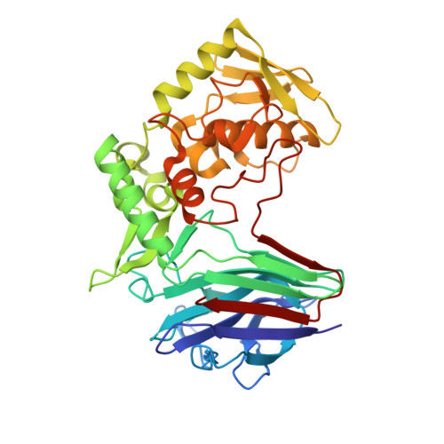 | |
UniProt | |||||
Find proteins for A0A8D3WIQ3 (Streptomyces pratensis (strain ATCC 33331 / IAF-45CD)) Explore A0A8D3WIQ3 Go to UniProtKB: A0A8D3WIQ3 | |||||
Entity Groups | |||||
| Sequence Clusters | 30% Identity50% Identity70% Identity90% Identity95% Identity100% Identity | ||||
| UniProt Group | A0A8D3WIQ3 | ||||
Sequence AnnotationsExpand | |||||
| |||||
| Length ( Å ) | Angle ( ˚ ) |
|---|---|
| a = 78.989 | α = 90 |
| b = 78.989 | β = 90 |
| c = 157.647 | γ = 120 |
| Software Name | Purpose |
|---|---|
| HKL-3000 | data collection |
| PHENIX | refinement |
| HKL-3000 | data reduction |
| HKL-3000 | data scaling |
| PHENIX | model building |
| PHENIX | phasing |
| Funding Organization | Location | Grant Number |
|---|---|---|
| National Natural Science Foundation of China (NSFC) | China | -- |
RCSB PDB (citation) is hosted by
RCSB PDB is a member of the

