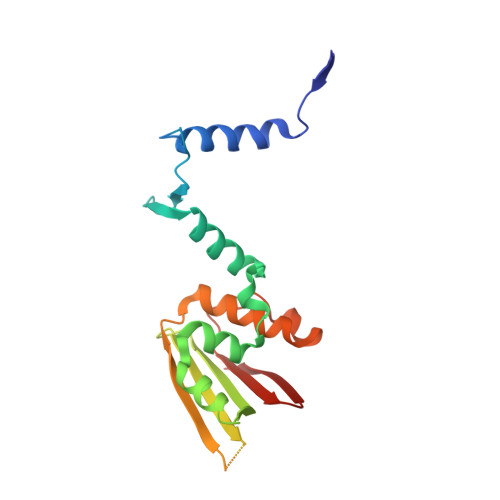
7UYX

Structure of bacteriophage PA1c gp2


Experimental Data Snapshot

  • Method: X-RAY DIFFRACTION
  • Resolution: 2.63 Å
  • R-Value Free: 0.300 
  • R-Value Work: 0.271 
  • R-Value Observed: 0.272 

wwPDB Validation   3D Report Full Report


This is version 1.3 of the entry. See complete history


Literature

Identification of the bacteriophage nucleus protein interaction network.

Enustun, E.Deep, A.Gu, Y.Nguyen, K.T.Chaikeeratisak, V.Armbruster, E.Ghassemian, M.Villa, E.Pogliano, J.Corbett, K.D.

(2023) Nat Struct Mol Biol 30: 1653-1662

  • DOI: https://doi.org/10.1038/s41594-023-01094-5
  • Primary Citation of Related Structures:  
    7UYX

  • PubMed Abstract: 

    In the arms race between bacteria and bacteriophages (phages), some large-genome jumbo phages have evolved a protein shell that encloses their replicating genome to protect it against host immune factors. By segregating the genome from the host cytoplasm, however, the 'phage nucleus' introduces the need to specifically translocate messenger RNA and proteins through the nuclear shell and to dock capsids on the shell for genome packaging. Here, we use proximity labeling and localization mapping to systematically identify proteins associated with the major nuclear shell protein chimallin (ChmA) and other distinctive structures assembled by these phages. We identify six uncharacterized nuclear-shell-associated proteins, one of which directly interacts with self-assembled ChmA. The structure and protein-protein interaction network of this protein, which we term ChmB, suggest that it forms pores in the ChmA lattice that serve as docking sites for capsid genome packaging and may also participate in messenger RNA and/or protein translocation.


  • Organizational Affiliation

    Department of Molecular Biology, University of California San Diego, La Jolla, CA, USA.


Macromolecules
Find similar proteins by:  (by identity cutoff)  |  3D Structure
Entity ID: 1
MoleculeChains Sequence LengthOrganismDetailsImage
Bacteriophage PA1C gp2
A, B, C, D
196Pseudomonas phage PA1CMutation(s): 0 
UniProt
Find proteins for A0A4D6BFJ2 (Pseudomonas phage PA1C)
Explore A0A4D6BFJ2 
Go to UniProtKB:  A0A4D6BFJ2
Entity Groups  
Sequence Clusters30% Identity50% Identity70% Identity90% Identity95% Identity100% Identity
UniProt GroupA0A4D6BFJ2
Sequence Annotations
Expand
  • Reference Sequence
Experimental Data & Validation

Experimental Data

  • Method: X-RAY DIFFRACTION
  • Resolution: 2.63 Å
  • R-Value Free: 0.300 
  • R-Value Work: 0.271 
  • R-Value Observed: 0.272 
  • Space Group: P 1 21 1
Unit Cell:
Length ( Å )Angle ( ˚ )
a = 60.955α = 90
b = 86.563β = 110.94
c = 71.879γ = 90
Software Package:
Software NamePurpose
PHENIXrefinement
XDSdata reduction
Aimlessdata scaling
PHASERphasing

Structure Validation

View Full Validation Report


Entry History & Funding Information

Deposition Data


Funding OrganizationLocationGrant Number
National Institutes of Health/National Institute of General Medical Sciences (NIH/NIGMS)United StatesR35 GM144121
National Institutes of Health/National Institute of General Medical Sciences (NIH/NIGMS)United StatesR01 GM129245

Revision History  (Full details and data files)

  • Version 1.0: 2023-05-10
    Type: Initial release
  • Version 1.1: 2023-09-06
    Changes: Data collection, Database references
  • Version 1.2: 2024-04-03
    Changes: Refinement description
  • Version 1.3: 2024-04-10
    Changes: Database references