7US5

X-ray crystal structure of GDP-D-glycero-D-manno-heptose 4,6-Dehydratase from Campylobacter jejuni


Experimental Data Snapshot

  • Method: X-RAY DIFFRACTION
  • Resolution: 2.10 Å
  • R-Value Free: 0.221 
  • R-Value Work: 0.169 
  • R-Value Observed: 0.172 

wwPDB Validation   3D Report Full Report


Ligand Structure Quality Assessment 


This is version 1.2 of the entry. See complete history


Literature

Reaction Mechanism and Three-Dimensional Structure of GDP-d-glycero-alpha-d-manno-heptose 4,6-Dehydratase from Campylobacter jejuni.

Xiang, D.F.Thoden, J.B.Ghosh, M.K.Holden, H.M.Raushel, F.M.

(2022) Biochemistry 61: 1313-1322

  • DOI: https://doi.org/10.1021/acs.biochem.2c00244
  • Primary Citation of Related Structures:  
    7US5

  • PubMed Abstract: 

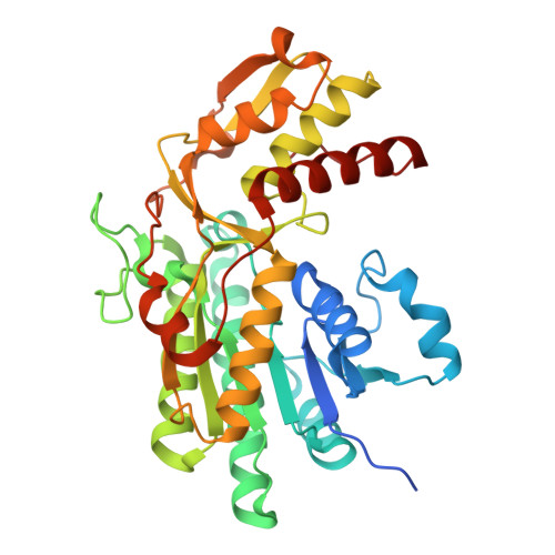
    Campylobacter jejuni is a human pathogen and a leading cause of food poisoning in the United States and Europe. Surrounding the outside of the bacterium is a carbohydrate coat known as the capsular polysaccharide. Various strains of C. jejuni have different sequences of unusual sugars and an assortment of decorations. Many of the serotypes have heptoses with differing stereochemical arrangements at C2 through C6. One of the many common modifications is a 6-deoxy-heptose that is formed by dehydration of GDP-d-glycero-α-d-manno-heptose to GDP-6-deoxy-4-keto-d-lyxo-heptose via the action of the enzyme GDP-d-glycero-α-d-manno-heptose 4,6-dehydratase. Herein, we report the biochemical and structural characterization of this enzyme from C. jejuni 81-176 (serotype HS:23/36). The enzyme was purified to homogeneity, and its three-dimensional structure was determined to a resolution of 2.1 Å. Kinetic analyses suggest that the reaction mechanism proceeds through the formation of a 4-keto intermediate followed by the loss of water from C5/C6. Based on the three-dimensional structure, it is proposed that oxidation of C4 is assisted by proton transfer from the hydroxyl group to the phenolate of Tyr-159 and hydride transfer to the tightly bound NAD + in the active site. Elimination of water at C5/C6 is most likely assisted by abstraction of the proton at C5 by Glu-136 and subsequent proton transfer to the hydroxyl at C6 via Ser-134 and Tyr-159. A bioinformatic analysis identified 19 additional 4,6-dehydratases from serotyped strains of C. jejuni that are 89-98% identical in the amino acid sequence, indicating that each of these strains should contain a 6-deoxy-heptose within their capsular polysaccharides.


  • Organizational Affiliation

    Department of Chemistry, Texas A&M University, College Station, Texas 77843, United States.


Macromolecules
Find similar proteins by:  (by identity cutoff)  |  3D Structure
Entity ID: 1
MoleculeChains Sequence LengthOrganismDetailsImage
GDP-D-GLYCERO-D-MANNO-HEPTOSE 4,6-DEHYDRATASE
A, B, C, D
365Campylobacter jejuniMutation(s): 0 
Gene Names: dmhABBR99_05330F1P94_08755HS23.12
UniProt
Find proteins for Q5M6Q7 (Campylobacter jejuni)
Explore Q5M6Q7 
Go to UniProtKB:  Q5M6Q7
Entity Groups  
Sequence Clusters30% Identity50% Identity70% Identity90% Identity95% Identity100% Identity
UniProt GroupQ5M6Q7
Sequence Annotations
Expand
  • Reference Sequence
Small Molecules
Ligands 3 Unique
IDChains Name / Formula / InChI Key2D Diagram3D Interactions
NAD (Subject of Investigation/LOI)
Query on NAD

Download Ideal Coordinates CCD File 
E [auth A],
H [auth B],
K [auth C],
P [auth D]
NICOTINAMIDE-ADENINE-DINUCLEOTIDE
C21 H27 N7 O14 P2
BAWFJGJZGIEFAR-NNYOXOHSSA-N
GDP (Subject of Investigation/LOI)
Query on GDP

Download Ideal Coordinates CCD File 
F [auth A],
I [auth B],
L [auth C],
Q [auth D]
GUANOSINE-5'-DIPHOSPHATE
C10 H15 N5 O11 P2
QGWNDRXFNXRZMB-UUOKFMHZSA-N
EDO
Query on EDO

Download Ideal Coordinates CCD File 
G [auth A]
J [auth B]
M [auth C]
N [auth C]
O [auth C]
G [auth A],
J [auth B],
M [auth C],
N [auth C],
O [auth C],
R [auth D]
1,2-ETHANEDIOL
C2 H6 O2
LYCAIKOWRPUZTN-UHFFFAOYSA-N
Experimental Data & Validation

Experimental Data

  • Method: X-RAY DIFFRACTION
  • Resolution: 2.10 Å
  • R-Value Free: 0.221 
  • R-Value Work: 0.169 
  • R-Value Observed: 0.172 
  • Space Group: P 21 21 2
Unit Cell:
Length ( Å )Angle ( ˚ )
a = 112.673α = 90
b = 182.95β = 90
c = 75.397γ = 90
Software Package:
Software NamePurpose
REFMACrefinement
PDB_EXTRACTdata extraction
SAINTdata reduction
SADABSdata scaling
MrBUMPphasing

Structure Validation

View Full Validation Report


Ligand Structure Quality Assessment 


Entry History & Funding Information

Deposition Data


Funding OrganizationLocationGrant Number
National Institutes of Health/National Institute of General Medical Sciences (NIH/NIGMS)United StatesGM 139428
National Institutes of Health/National Institute of General Medical Sciences (NIH/NIGMS)United StatesGM 122825
National Institutes of Health/National Institute of General Medical Sciences (NIH/NIGMS)United StatesGM 134643

Revision History  (Full details and data files)

  • Version 1.0: 2022-07-06
    Type: Initial release
  • Version 1.1: 2022-07-13
    Changes: Database references
  • Version 1.2: 2023-10-18
    Changes: Data collection, Refinement description