


Crystal Structure of Siderophore Binding Protein FatB from Desulfitobacterium hafniense
Kim, Y., Patel, H.P., Nordquist, K.A., Schaab, K.M., Sha, J., Babnigg, G., Bond, A.H., Joachimiak, A., Midwest Center for Structural GenomicsTo be published.
Experimental Data Snapshot
Entity ID: 1 | |||||
|---|---|---|---|---|---|
| Molecule | Chains | Sequence Length | Organism | Details | Image |
| Ferric-anguibactin-binding protein FatB | 314 | Desulfitobacterium hafniense DCB-2 | Mutation(s): 0 Gene Names: Dhaf_2916 | 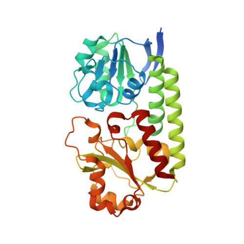 | |
UniProt | |||||
Find proteins for Q24WN9 (Desulfitobacterium hafniense (strain Y51)) Explore Q24WN9 Go to UniProtKB: Q24WN9 | |||||
Entity Groups | |||||
| Sequence Clusters | 30% Identity50% Identity70% Identity90% Identity95% Identity100% Identity | ||||
| UniProt Group | Q24WN9 | ||||
Sequence AnnotationsExpand | |||||
| |||||
| Ligands 5 Unique | |||||
|---|---|---|---|---|---|
| ID | Chains | Name / Formula / InChI Key | 2D Diagram | 3D Interactions | |
| DBS (Subject of Investigation/LOI) Query on DBS | B [auth A], C [auth A] | 2-(2,3-DIHYDROXY-BENZOYLAMINO)-3-HYDROXY-PROPIONIC ACID C10 H11 N O6 VDTYHTVHFIIEIL-LURJTMIESA-N |  | ||
| GOL Query on GOL | D [auth A] | GLYCEROL C3 H8 O3 PEDCQBHIVMGVHV-UHFFFAOYSA-N |  | ||
| EDO Query on EDO | E [auth A], F [auth A], G [auth A] | 1,2-ETHANEDIOL C2 H6 O2 LYCAIKOWRPUZTN-UHFFFAOYSA-N |  | ||
| FE Query on FE | H [auth A] | FE (III) ION Fe VTLYFUHAOXGGBS-UHFFFAOYSA-N |  | ||
| CL Query on CL | I [auth A], J [auth A] | CHLORIDE ION Cl VEXZGXHMUGYJMC-UHFFFAOYSA-M |  | ||
| Length ( Å ) | Angle ( ˚ ) |
|---|---|
| a = 78.884 | α = 90 |
| b = 78.884 | β = 90 |
| c = 122.402 | γ = 120 |
| Software Name | Purpose |
|---|---|
| PHENIX | refinement |
| HKL-3000 | data reduction |
| HKL-3000 | data scaling |
| HKL-3000 | phasing |
| MOLREP | phasing |
| Funding Organization | Location | Grant Number |
|---|---|---|
| National Institutes of Health/National Institute of General Medical Sciences (NIH/NIGMS) | United States | -- |
RCSB PDB (citation) is hosted by
RCSB PDB is a member of the

