7S43

Crystal structure of an N-acetyltransferase, C80T mutant, from Helicobacter pullorum in the presence of Coenzyme A and dTDP-3-amino-3,6-dideoxy-D-glucose


Experimental Data Snapshot

  • Method: X-RAY DIFFRACTION
  • Resolution: 1.70 Å
  • R-Value Free: 0.192 
  • R-Value Work: 0.159 
  • R-Value Observed: 0.161 

wwPDB Validation   3D Report Full Report


Ligand Structure Quality Assessment 


This is version 1.3 of the entry. See complete history


Literature

Biochemical investigation of an N-acetyltransferase from Helicobacter pullorum.

Griffiths, W.A.Spencer, K.D.Thoden, J.B.Holden, H.M.

(2021) Protein Sci 30: 2418-2432

  • DOI: https://doi.org/10.1002/pro.4207
  • Primary Citation of Related Structures:  
    7S3U, 7S3W, 7S41, 7S42, 7S43, 7S44, 7S45

  • PubMed Abstract: 

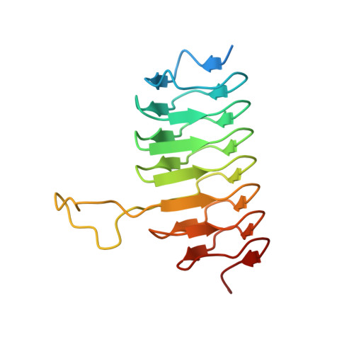
    N-acetylated sugars are often found, for example, on the lipopolysaccharides of Gram-negative bacteria, on the S-layers of Gram-positive bacteria, and on the capsular polysaccharides. Key enzymes involved in their biosynthesis are the sugar N-acetyltransferases. Here, we describe a structural and functional analysis of one such enzyme from Helicobacter pullorum, an emerging pathogen that may be associated with gastroenteritis and gallbladder and liver diseases. For this analysis, the gene BA919-RS02330 putatively encoding an N-acetyltransferase was cloned, and the corresponding protein was expressed and purified. A kinetic analysis demonstrated that the enzyme utilizes dTDP-3-amino-3,6-dideoxy-d-glucose as a substrate as well as dTDP-3-amino-3,6-dideoxy-d-galactose, albeit at a reduced rate. In addition to this kinetic analysis, a similar enzyme from Helicobacter bilis was cloned and expressed, and its kinetic parameters were determined. Seven X-ray crystallographic structures of various complexes of the H. pullorum wild-type enzyme (or the C80T variant) were determined to resolutions of 1.7 Å or higher. The overall molecular architecture of the H. pullorum N-acetyltransferase places it into the Class II left-handed-β-helix superfamily (LβH). Taken together, the data presented herein suggest that 3-acetamido-3,6-dideoxy-d-glucose (or the galactose derivative) is found on either the H. pullorum O-antigen or in another of its complex glycoconjugates. A BLAST search suggests that more than 50 non-pylori Helicobacter spp. have genes encoding N-acetyltransferases. Given that there is little information concerning the complex glycans in non-pylori Helicobacter spp. and considering their zoonotic potential, our results provide new biochemical insight into these pathogens.


  • Organizational Affiliation

    Department of Biochemistry, University of Wisconsin, Madison, Wisconsin, USA.


Macromolecules
Find similar proteins by:  (by identity cutoff)  |  3D Structure
Entity ID: 1
MoleculeChains Sequence LengthOrganismDetailsImage
N-acetyltransferase165Helicobacter pullorumMutation(s): 0 
Gene Names: BA919_rs02330
Entity Groups  
Sequence Clusters30% Identity50% Identity70% Identity90% Identity95% Identity100% Identity
Sequence Annotations
Expand
  • Reference Sequence
Small Molecules
Ligands 4 Unique
IDChains Name / Formula / InChI Key2D Diagram3D Interactions
COA (Subject of Investigation/LOI)
Query on COA

Download Ideal Coordinates CCD File 
C [auth A]COENZYME A
C21 H36 N7 O16 P3 S
RGJOEKWQDUBAIZ-IBOSZNHHSA-N
T3Q (Subject of Investigation/LOI)
Query on T3Q

Download Ideal Coordinates CCD File 
B [auth A][(3R,4S,5S,6R)-4-amino-3,5-dihydroxy-6-methyloxan-2-yl][hydroxy-[[(2R,3S,5R)-3-hydroxy-5-(5-methyl-2,4-dioxopyrimidin-1-yl)oxolan-2-yl]methoxy]phosphoryl] hydrogen phosphate
C16 H27 N3 O14 P2
KVYJLJOGNUNRJK-HALQBZCBSA-N
EDO
Query on EDO

Download Ideal Coordinates CCD File 
D [auth A]1,2-ETHANEDIOL
C2 H6 O2
LYCAIKOWRPUZTN-UHFFFAOYSA-N
NA
Query on NA

Download Ideal Coordinates CCD File 
E [auth A]SODIUM ION
Na
FKNQFGJONOIPTF-UHFFFAOYSA-N
Experimental Data & Validation

Experimental Data

  • Method: X-RAY DIFFRACTION
  • Resolution: 1.70 Å
  • R-Value Free: 0.192 
  • R-Value Work: 0.159 
  • R-Value Observed: 0.161 
  • Space Group: I 2 3
Unit Cell:
Length ( Å )Angle ( ˚ )
a = 102.844α = 90
b = 102.844β = 90
c = 102.844γ = 90
Software Package:
Software NamePurpose
REFMACrefinement
PDB_EXTRACTdata extraction
SAINTdata reduction
SADABSdata scaling
REFMACphasing

Structure Validation

View Full Validation Report


Ligand Structure Quality Assessment 


Entry History & Funding Information

Deposition Data


Funding OrganizationLocationGrant Number
National Institutes of Health/National Institute of General Medical Sciences (NIH/NIGMS)United StatesR35 GM134643

Revision History  (Full details and data files)

  • Version 1.0: 2021-09-22
    Type: Initial release
  • Version 1.1: 2021-11-10
    Changes: Database references
  • Version 1.2: 2021-12-08
    Changes: Database references
  • Version 1.3: 2023-10-18
    Changes: Data collection, Refinement description