


Crystal structure of Triosephosphate isomerase from Ktedonobacter racemifer
Vickers, C.J., Patrick, W.M., Fraga, D.To be published.
Experimental Data Snapshot
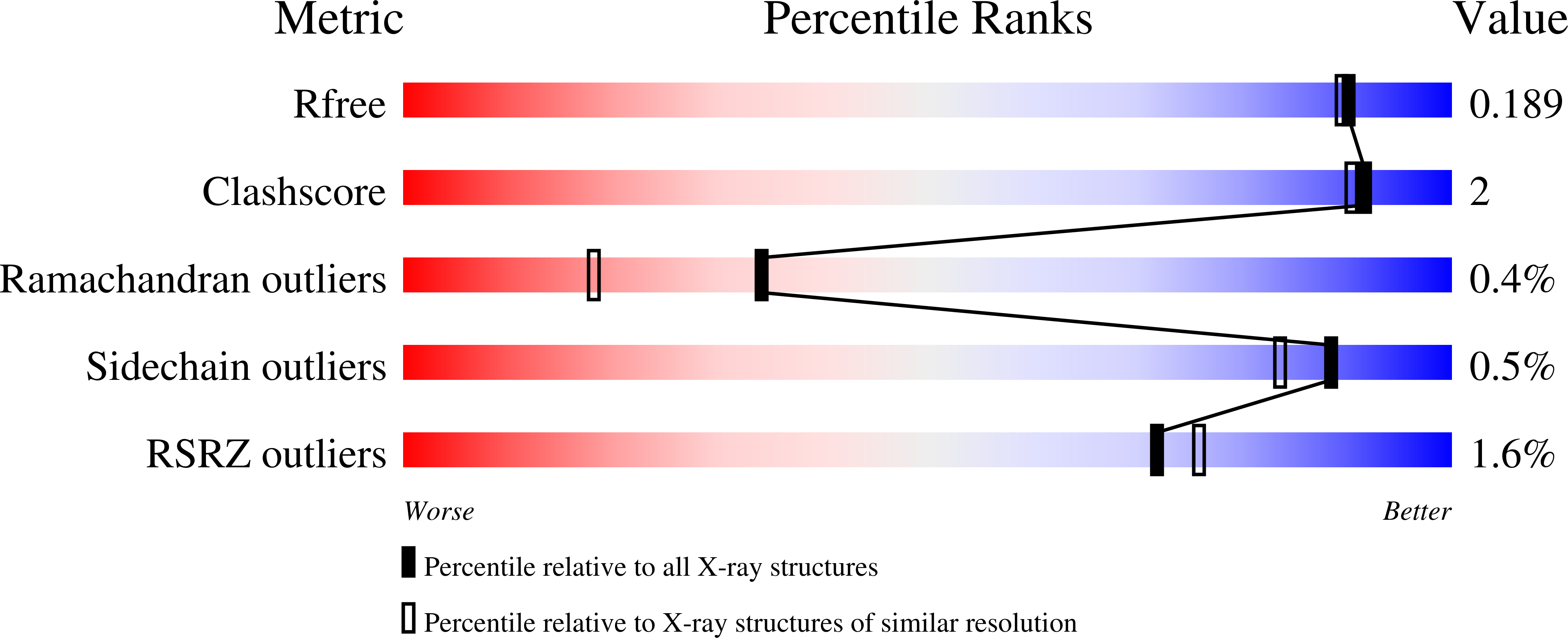
wwPDB Validation 3D Report Full Report
Entity ID: 1 | |||||
|---|---|---|---|---|---|
| Molecule | Chains | Sequence Length | Organism | Details | Image |
| Triosephosphate isomerase | 269 | Ktedonobacter racemifer | Mutation(s): 0 Gene Names: tpiA, Krac_9127 EC: 5.3.1.1 |  | |
UniProt | |||||
Find proteins for D6TR79 (Ktedonobacter racemifer DSM 44963) Explore D6TR79 Go to UniProtKB: D6TR79 | |||||
Entity Groups | |||||
| Sequence Clusters | 30% Identity50% Identity70% Identity90% Identity95% Identity100% Identity | ||||
| UniProt Group | D6TR79 | ||||
Sequence AnnotationsExpand | |||||
| |||||
| Ligands 1 Unique | |||||
|---|---|---|---|---|---|
| ID | Chains | Name / Formula / InChI Key | 2D Diagram | 3D Interactions | |
| NO3 (Subject of Investigation/LOI) Query on NO3 | B [auth A], C [auth A], D [auth A] | NITRATE ION N O3 NHNBFGGVMKEFGY-UHFFFAOYSA-N |  | ||
| Length ( Å ) | Angle ( ˚ ) |
|---|---|
| a = 64.58 | α = 90 |
| b = 54.741 | β = 115.196 |
| c = 74.843 | γ = 90 |
| Software Name | Purpose |
|---|---|
| PHENIX | refinement |
| MOSFLM | data reduction |
| Aimless | data scaling |
| PHASER | phasing |
| Funding Organization | Location | Grant Number |
|---|---|---|
| Royal Society of New Zealand | New Zealand | -- |
RCSB PDB (citation) is hosted by
RCSB PDB is a member of the

