7RAG

Structure of the CwlD amidase from Clostridioides difficile in complex with the GerS lipoprotein

  • Classification: HYDROLASE
  • Organism(s): Clostridioides difficile
  • Expression System: Escherichia coli
  • Mutation(s): No 

  • Deposited: 2021-07-01 Released: 2021-09-08 
  • Deposition Author(s): Eckenroth, B.E., Doublie, S.
  • Funding Organization(s): National Institutes of Health/National Institute of General Medical Sciences (NIH/NIGMS), National Institutes of Health/National Cancer Institute (NIH/NCI)

Experimental Data Snapshot

  • Method: X-RAY DIFFRACTION
  • Resolution: 2.40 Å
  • R-Value Free: 0.249 
  • R-Value Work: 0.203 
  • R-Value Observed: 0.208 

wwPDB Validation   3D Report Full Report


This is version 1.1 of the entry. See complete history


Literature

A lipoprotein allosterically activates the CwlD amidase during Clostridioides difficile spore formation.

Alves Feliciano, C.Eckenroth, B.E.Diaz, O.R.Doublie, S.Shen, A.

(2021) PLoS Genet 17: e1009791-e1009791

  • DOI: https://doi.org/10.1371/journal.pgen.1009791
  • Primary Citation of Related Structures:  
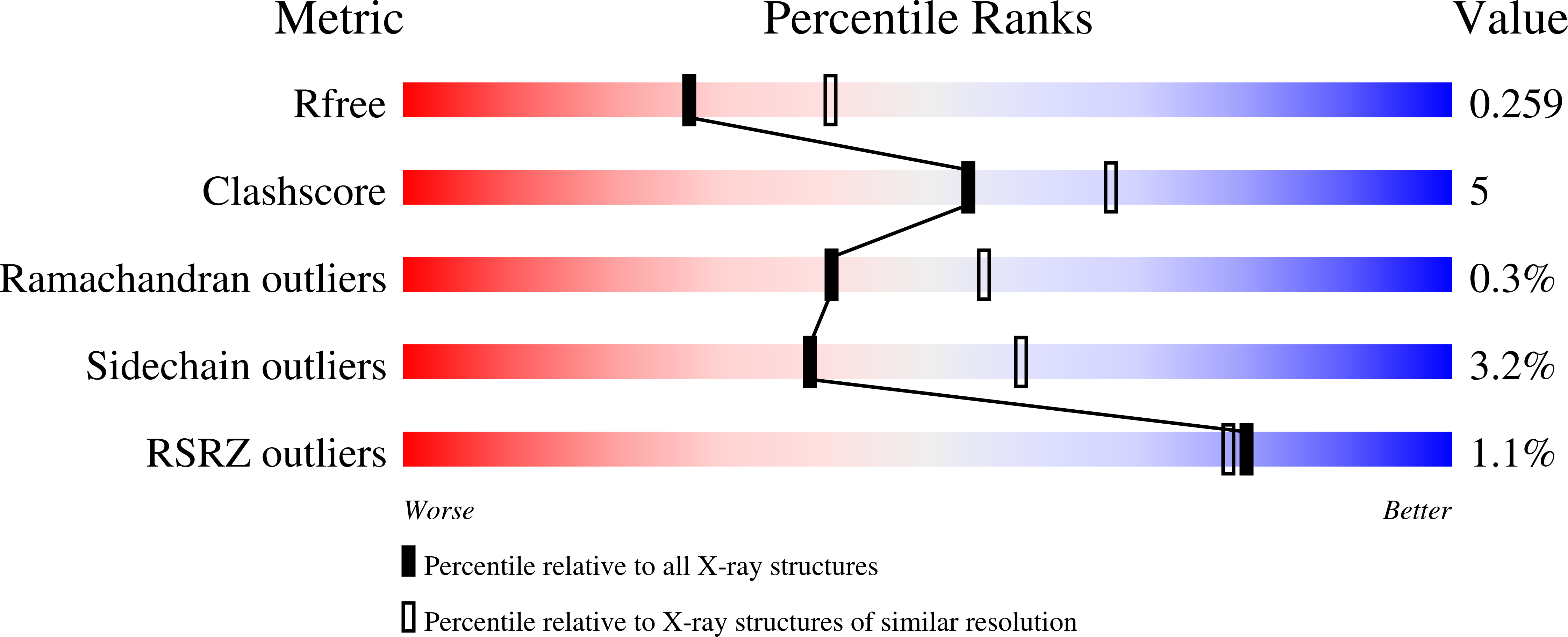
    7RAG

  • PubMed Abstract: 

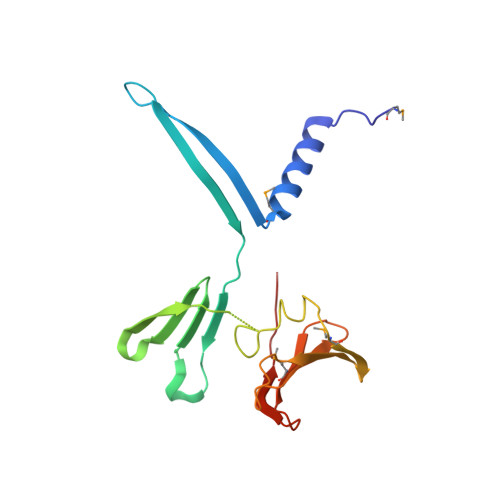
    Spore-forming pathogens like Clostridioides difficile depend on germination to initiate infection. During gemination, spores must degrade their cortex layer, which is a thick, protective layer of modified peptidoglycan. Cortex degradation depends on the presence of the spore-specific peptidoglycan modification, muramic-∂-lactam (MAL), which is specifically recognized by cortex lytic enzymes. In C. difficile, MAL production depends on the CwlD amidase and its binding partner, the GerS lipoprotein. To gain insight into how GerS regulates CwlD activity, we solved the crystal structure of the CwlD:GerS complex. In this structure, a GerS homodimer is bound to two CwlD monomers such that the CwlD active sites are exposed. Although CwlD structurally resembles amidase_3 family members, we found that CwlD does not bind Zn2+ stably on its own, unlike previously characterized amidase_3 enzymes. Instead, GerS binding to CwlD promotes CwlD binding to Zn2+, which is required for its catalytic mechanism. Thus, in determining the first structure of an amidase bound to its regulator, we reveal stabilization of Zn2+ co-factor binding as a novel mechanism for regulating bacterial amidase activity. Our results further suggest that allosteric regulation by binding partners may be a more widespread mode for regulating bacterial amidase activity than previously thought.


  • Organizational Affiliation

    Department of Molecular Biology and Microbiology, Tufts University School of Medicine, Boston, Massachusetts, United States of America.


Macromolecules
Find similar proteins by:  (by identity cutoff)  |  3D Structure
Entity ID: 1
MoleculeChains Sequence LengthOrganismDetailsImage
Lipoprotein175Clostridioides difficileMutation(s): 0 
Gene Names: 
UniProt
Find proteins for Q180W3 (Clostridioides difficile (strain 630))
Explore Q180W3 
Go to UniProtKB:  Q180W3
Entity Groups  
Sequence Clusters30% Identity50% Identity70% Identity90% Identity95% Identity100% Identity
UniProt GroupQ180W3
Sequence Annotations
Expand
  • Reference Sequence
Find similar proteins by:  (by identity cutoff)  |  3D Structure
Entity ID: 2
MoleculeChains Sequence LengthOrganismDetailsImage
Germination-specific N-acetylmuramoyl-L-alanine amidase, Autolysin217Clostridioides difficileMutation(s): 0 
Gene Names: cwlDCD630_01060
EC: 3.5.1.28
UniProt
Find proteins for Q18CJ4 (Clostridioides difficile (strain 630))
Explore Q18CJ4 
Go to UniProtKB:  Q18CJ4
Entity Groups  
Sequence Clusters30% Identity50% Identity70% Identity90% Identity95% Identity100% Identity
UniProt GroupQ18CJ4
Sequence Annotations
Expand
  • Reference Sequence
Small Molecules
Modified Residues  1 Unique
IDChains TypeFormula2D DiagramParent
MSE
Query on MSE
A
L-PEPTIDE LINKINGC5 H11 N O2 SeMET
Experimental Data & Validation

Experimental Data

  • Method: X-RAY DIFFRACTION
  • Resolution: 2.40 Å
  • R-Value Free: 0.249 
  • R-Value Work: 0.203 
  • R-Value Observed: 0.208 
  • Space Group: P 65 2 2
Unit Cell:
Length ( Å )Angle ( ˚ )
a = 105.185α = 90
b = 105.185β = 90
c = 154.771γ = 120
Software Package:
Software NamePurpose
PROTEUM PLUSdata reduction
PROTEUM PLUSdata scaling
SHARPphasing
PHENIXrefinement
PDB_EXTRACTdata extraction

Structure Validation

View Full Validation Report


Entry History & Funding Information

Deposition Data


Funding OrganizationLocationGrant Number
National Institutes of Health/National Institute of General Medical Sciences (NIH/NIGMS)United StatesGM140361
National Institutes of Health/National Cancer Institute (NIH/NCI)United StatesCA233185

Revision History  (Full details and data files)

  • Version 1.0: 2021-09-08
    Type: Initial release
  • Version 1.1: 2021-10-06
    Changes: Database references