7MDH

STRUCTURAL BASIS FOR LIGHT ACITVATION OF A CHLOROPLAST ENZYME. THE STRUCTURE OF SORGHUM NADP-MALATE DEHYDROGENASE IN ITS OXIDIZED FORM


Experimental Data Snapshot

  • Method: X-RAY DIFFRACTION
  • Resolution: 2.40 Å
  • R-Value Free: 0.271 
  • R-Value Work: 0.221 

wwPDB Validation   3D Report Full Report


This is version 1.4 of the entry. See complete history


Literature

Structural basis for light activation of a chloroplast enzyme: the structure of sorghum NADP-malate dehydrogenase in its oxidized form.

Johansson, K.Ramaswamy, S.Saarinen, M.Lemaire-Chamley, M.Issakidis-Bourguet, E.Miginiac-Maslow, M.Eklund, H.

(1999) Biochemistry 38: 4319-4326

  • DOI: https://doi.org/10.1021/bi982876c
  • Primary Citation of Related Structures:  
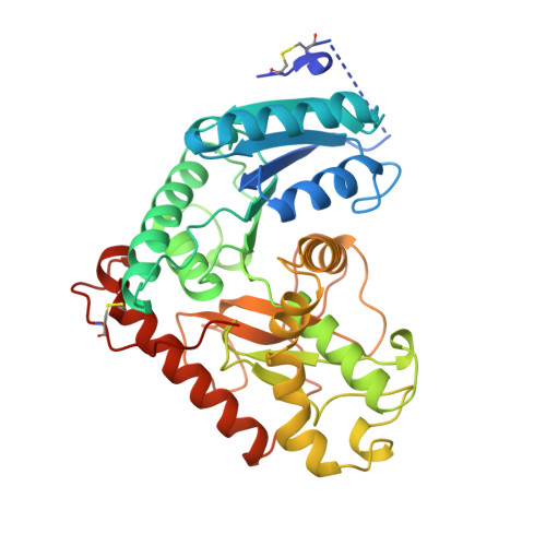
    7MDH

  • PubMed Abstract: 

    Some key chloroplast enzymes are activated by light via a ferredoxin-thioredoxin reduction system which reduces disulfide bridges in the enzymes. We describe for the first time the structural basis for the redox activation of a chloroplast enzyme, the NADP-dependent malate dehydrogenase (MDH) from Sorghum vulgare whose structure has been determined and refined at 2.4 A resolution. In addition to the normal structural components of MDHs, the enzyme exhibits extensions at both the N- and C-termini, each of which contains a regulatory disulfide bridge which must be reduced for activation. The N-terminal disulfide motif is inserted in a cleft between the two subunits of the dimer, thereby locking the domains in each subunit. The C-terminal disulfide keeps the C-terminal residues tight to the enzyme surface and blocks access to the active site. Reduction of the N-terminal disulfide would release the stopper between the domains and give the enzyme the necessary flexibility. Simultaneous reduction of the C-terminal disulfide would free the C-terminal residues from binding to the enzyme and make the active site accessible.


  • Organizational Affiliation

    Department of Molecular Biology, Swedish University of Agricultural Sciences, Uppsala.


Macromolecules
Find similar proteins by:  (by identity cutoff)  |  3D Structure
Entity ID: 1
MoleculeChains Sequence LengthOrganismDetailsImage
PROTEIN (MALATE DEHYDROGENASE)
A, B, C, D
375Sorghum bicolorMutation(s): 0 
EC: 1.1.1.82
UniProt
Find proteins for P17606 (Sorghum bicolor)
Explore P17606 
Go to UniProtKB:  P17606
Entity Groups  
Sequence Clusters30% Identity50% Identity70% Identity90% Identity95% Identity100% Identity
UniProt GroupP17606
Sequence Annotations
Expand
  • Reference Sequence
Small Molecules
Ligands 1 Unique
IDChains Name / Formula / InChI Key2D Diagram3D Interactions
ZN
Query on ZN

Download Ideal Coordinates CCD File 
E [auth A]
F [auth A]
G [auth A]
H [auth A]
I [auth B]
E [auth A],
F [auth A],
G [auth A],
H [auth A],
I [auth B],
J [auth B],
K [auth B],
L [auth B],
M [auth B],
N [auth C],
O [auth C],
P [auth C],
Q [auth C],
R [auth C],
S [auth D],
T [auth D],
U [auth D]
ZINC ION
Zn
PTFCDOFLOPIGGS-UHFFFAOYSA-N
Experimental Data & Validation

Experimental Data

  • Method: X-RAY DIFFRACTION
  • Resolution: 2.40 Å
  • R-Value Free: 0.271 
  • R-Value Work: 0.221 
  • Space Group: C 2 2 21
Unit Cell:
Length ( Å )Angle ( ˚ )
a = 145.424α = 90
b = 153.944β = 90
c = 160.042γ = 90
Software Package:
Software NamePurpose
DENZOdata reduction
SCALEPACKdata scaling
AMoREphasing
CNSrefinement

Structure Validation

View Full Validation Report


Entry History 

Deposition Data

Revision History  (Full details and data files)

  • Version 1.0: 1999-06-04
    Type: Initial release
  • Version 1.1: 2007-10-16
    Changes: Version format compliance
  • Version 1.2: 2011-07-13
    Changes: Derived calculations, Version format compliance
  • Version 1.3: 2018-03-07
    Changes: Data collection
  • Version 1.4: 2023-09-20
    Changes: Data collection, Database references, Derived calculations, Refinement description