7LLJ

Inhibitory immune receptor protein complex


Experimental Data Snapshot

  • Method: X-RAY DIFFRACTION
  • Resolution: 3.15 Å
  • R-Value Free: 0.276 
  • R-Value Work: 0.240 
  • R-Value Observed: 0.248 

wwPDB Validation   3D Report Full Report


Ligand Structure Quality Assessment 


This is version 1.2 of the entry. See complete history


Literature

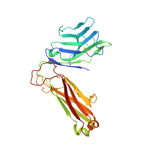
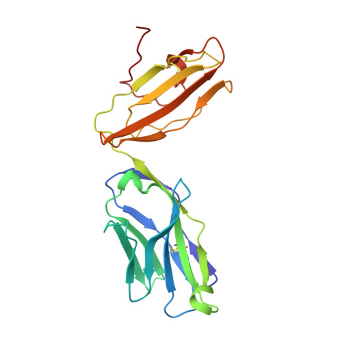
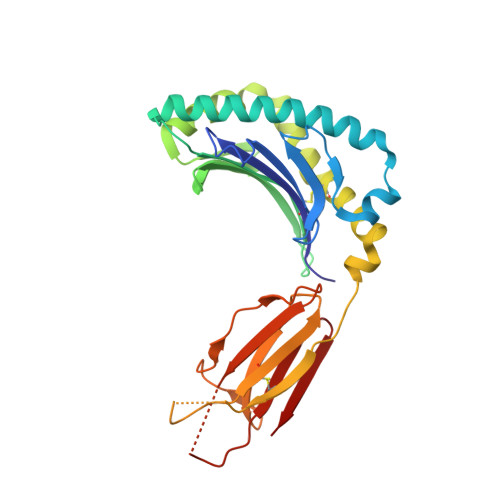
Recognition of the antigen-presenting molecule MR1 by a V delta 3 + gamma delta T cell receptor.

Rice, M.T.von Borstel, A.Chevour, P.Awad, W.Howson, L.J.Littler, D.R.Gherardin, N.A.Le Nours, J.Giles, E.M.Berry, R.Godfrey, D.I.Davey, M.S.Rossjohn, J.Gully, B.S.

(2021) Proc Natl Acad Sci U S A 118

  • DOI: https://doi.org/10.1073/pnas.2110288118
  • Primary Citation of Related Structures:  
    7LLI, 7LLJ

  • PubMed Abstract: 

    Unlike conventional αβ T cells, γδ T cells typically recognize nonpeptide ligands independently of major histocompatibility complex (MHC) restriction. Accordingly, the γδ T cell receptor (TCR) can potentially recognize a wide array of ligands; however, few ligands have been described to date. While there is a growing appreciation of the molecular bases underpinning variable (V)δ1 + and Vδ2 + γδ TCR-mediated ligand recognition, the mode of Vδ3 + TCR ligand engagement is unknown. MHC class I-related protein, MR1, presents vitamin B metabolites to αβ T cells known as mucosal-associated invariant T cells, diverse MR1-restricted T cells, and a subset of human γδ T cells. Here, we identify Vδ1/2 - γδ T cells in the blood and duodenal biopsy specimens of children that showed metabolite-independent binding of MR1 tetramers. Characterization of one Vδ3Vγ8 TCR clone showed MR1 reactivity was independent of the presented antigen. Determination of two Vδ3Vγ8 TCR-MR1-antigen complex structures revealed a recognition mechanism by the Vδ3 TCR chain that mediated specific contacts to the side of the MR1 antigen-binding groove, representing a previously uncharacterized MR1 docking topology. The binding of the Vδ3 + TCR to MR1 did not involve contacts with the presented antigen, providing a basis for understanding its inherent MR1 autoreactivity. We provide molecular insight into antigen-independent recognition of MR1 by a Vδ3 + γδ TCR that strengthens an emerging paradigm of antibody-like ligand engagement by γδ TCRs.


  • Organizational Affiliation

    Infection and Immunity Program, Biomedicine Discovery Institute, Monash University, Clayton, VIC 3800, Australia.


Macromolecules
Find similar proteins by:  (by identity cutoff)  |  3D Structure
Entity ID: 1
MoleculeChains Sequence LengthOrganismDetailsImage
T cell receptor gamma variable 8A [auth K],
E [auth A]
245Homo sapiensMutation(s): 0 
Gene Names: TRGV8
Entity Groups  
Sequence Clusters30% Identity50% Identity70% Identity90% Identity95% Identity100% Identity
Sequence Annotations
Expand
  • Reference Sequence
Find similar proteins by:  (by identity cutoff)  |  3D Structure
Entity ID: 2
MoleculeChains Sequence LengthOrganismDetailsImage
T cell receptor delta variable 3B [auth L],
F [auth B]
214Homo sapiensMutation(s): 0 
Gene Names: TRDV3
Entity Groups  
Sequence Clusters30% Identity50% Identity70% Identity90% Identity95% Identity100% Identity
Sequence Annotations
Expand
  • Reference Sequence
Find similar proteins by:  (by identity cutoff)  |  3D Structure
Entity ID: 3
MoleculeChains Sequence LengthOrganismDetailsImage
Major histocompatibility complex class I-related gene proteinC,
G [auth E]
271Homo sapiensMutation(s): 0 
Gene Names: MR1
UniProt & NIH Common Fund Data Resources
Find proteins for Q95460 (Homo sapiens)
Explore Q95460 
Go to UniProtKB:  Q95460
PHAROS:  Q95460
GTEx:  ENSG00000153029 
Entity Groups  
Sequence Clusters30% Identity50% Identity70% Identity90% Identity95% Identity100% Identity
UniProt GroupQ95460
Sequence Annotations
Expand
  • Reference Sequence
Find similar proteins by:  (by identity cutoff)  |  3D Structure
Entity ID: 4
MoleculeChains Sequence LengthOrganismDetailsImage
Beta-2-microglobulinD,
H [auth F]
100Homo sapiensMutation(s): 0 
Gene Names: B2MCDABP0092HDCMA22P
UniProt & NIH Common Fund Data Resources
Find proteins for P61769 (Homo sapiens)
Explore P61769 
Go to UniProtKB:  P61769
PHAROS:  P61769
GTEx:  ENSG00000166710 
Entity Groups  
Sequence Clusters30% Identity50% Identity70% Identity90% Identity95% Identity100% Identity
UniProt GroupP61769
Sequence Annotations
Expand
  • Reference Sequence
Small Molecules
Ligands 1 Unique
IDChains Name / Formula / InChI Key2D Diagram3D Interactions
30W (Subject of Investigation/LOI)
Query on 30W

Download Ideal Coordinates CCD File 
I [auth C],
J [auth E]
N-(6-formyl-4-oxo-3,4-dihydropteridin-2-yl)acetamide
C9 H7 N5 O3
DDBCPKAHJKOGKK-UHFFFAOYSA-N
Experimental Data & Validation

Experimental Data

  • Method: X-RAY DIFFRACTION
  • Resolution: 3.15 Å
  • R-Value Free: 0.276 
  • R-Value Work: 0.240 
  • R-Value Observed: 0.248 
  • Space Group: H 3
Unit Cell:
Length ( Å )Angle ( ˚ )
a = 297.549α = 90
b = 297.549β = 90
c = 120.371γ = 120
Software Package:
Software NamePurpose
PHENIXrefinement
Cootmodel building
MOSFLMdata reduction
Aimlessdata scaling
PHENIXphasing

Structure Validation

View Full Validation Report


Ligand Structure Quality Assessment 


Entry History & Funding Information

Deposition Data


Funding OrganizationLocationGrant Number
Australian Research Council (ARC)AustraliaFL160100049
Australian Research Council (ARC)AustraliaDP200103462

Revision History  (Full details and data files)

  • Version 1.0: 2021-11-17
    Type: Initial release
  • Version 1.1: 2021-12-08
    Changes: Database references
  • Version 1.2: 2023-10-18
    Changes: Data collection, Refinement description