


Crystal Structure of the RNA binding domain of Threonyl-tRNA synthetase from Cryptosporidium parvum Iowa II
Abendroth, J., Dranow, D.M., Lorimer, D.D., Horanyi, P.S., Edwards, T.E.To be published.
Experimental Data Snapshot
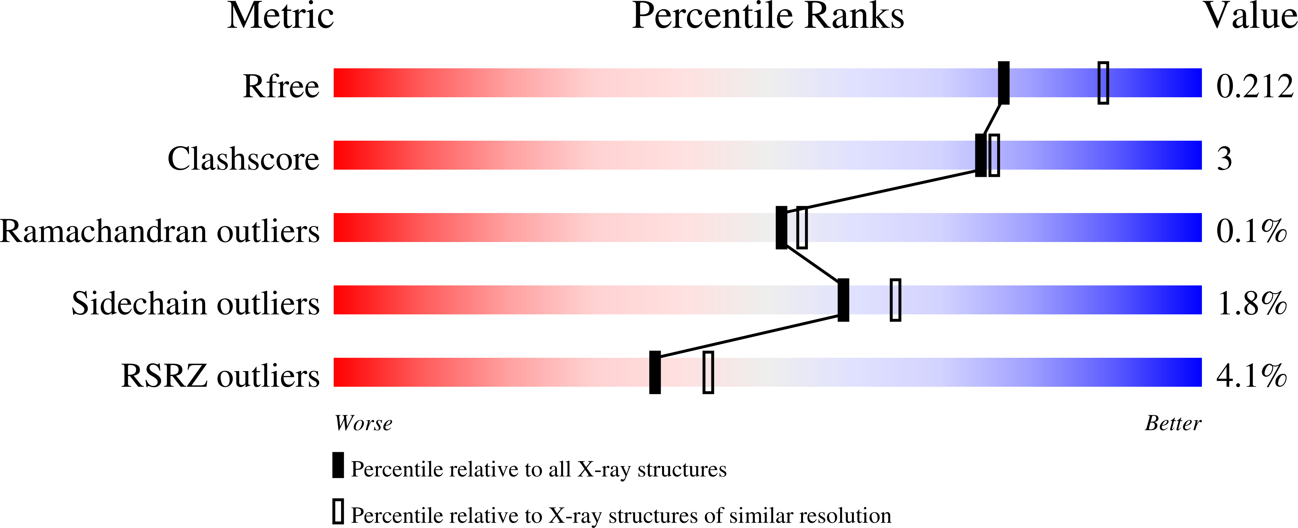
wwPDB Validation 3D Report Full Report
Entity ID: 1 | |||||
|---|---|---|---|---|---|
| Molecule | Chains | Sequence Length | Organism | Details | Image |
| Threonyl-tRNA synthetase | 442 | Cryptosporidium parvum Iowa II | Mutation(s): 0 Gene Names: cgd7_1710 EC: 6.1.1.3 | 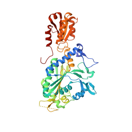 | |
UniProt | |||||
Find proteins for Q5CYN0 (Cryptosporidium parvum (strain Iowa II)) Explore Q5CYN0 Go to UniProtKB: Q5CYN0 | |||||
Entity Groups | |||||
| Sequence Clusters | 30% Identity50% Identity70% Identity90% Identity95% Identity100% Identity | ||||
| UniProt Group | Q5CYN0 | ||||
Sequence AnnotationsExpand | |||||
| |||||
| Ligands 2 Unique | |||||
|---|---|---|---|---|---|
| ID | Chains | Name / Formula / InChI Key | 2D Diagram | 3D Interactions | |
| ZN Query on ZN | E [auth A], G [auth B], I [auth C], J [auth D] | ZINC ION Zn PTFCDOFLOPIGGS-UHFFFAOYSA-N |  | ||
| CA Query on CA | F [auth A], H [auth B] | CALCIUM ION Ca BHPQYMZQTOCNFJ-UHFFFAOYSA-N |  | ||
| Length ( Å ) | Angle ( ˚ ) |
|---|---|
| a = 65.73 | α = 91.585 |
| b = 67.45 | β = 97.53 |
| c = 143.18 | γ = 120.777 |
| Software Name | Purpose |
|---|---|
| XDS | data reduction |
| XSCALE | data scaling |
| PHENIX | refinement |
| PDB_EXTRACT | data extraction |
| MoRDa | phasing |
| PHENIX | model building |
| Coot | model building |
RCSB PDB (citation) is hosted by
RCSB PDB is a member of the

