7KH2

Structure of N-citrylornithine decarboxylase bound with PLP


Experimental Data Snapshot

  • Method: X-RAY DIFFRACTION
  • Resolution: 2.05 Å
  • R-Value Free: 0.199 
  • R-Value Work: 0.174 
  • R-Value Observed: 0.174 

wwPDB Validation   3D Report Full Report


Ligand Structure Quality Assessment 


This is version 1.2 of the entry. See complete history


Literature

Alternative pathways utilize or circumvent putrescine for biosynthesis of putrescine-containing rhizoferrin.

Li, B.Deng, X.Kim, S.H.Buhrow, L.Tomchick, D.R.Phillips, M.A.Michael, A.J.

(2020) J Biol Chem 296: 100146-100146

  • DOI: https://doi.org/10.1074/jbc.RA120.016738
  • Primary Citation of Related Structures:  
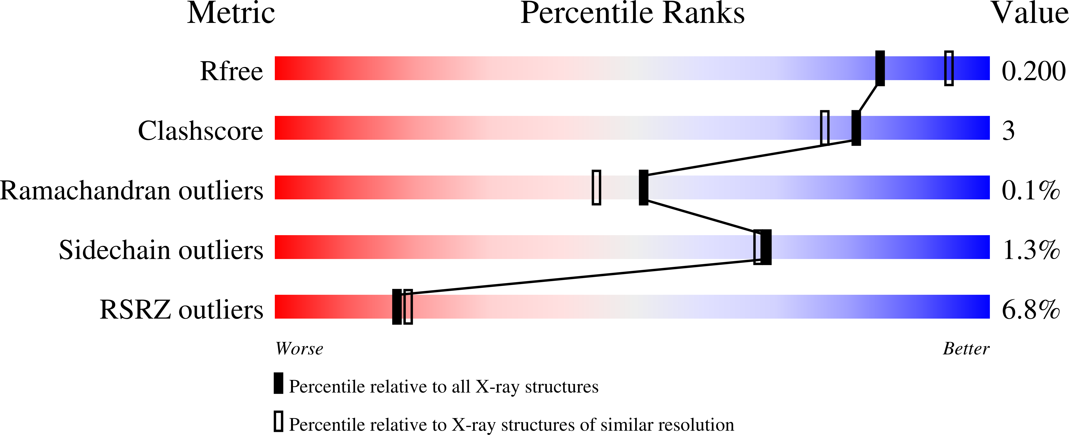
    7KH2

  • PubMed Abstract: 

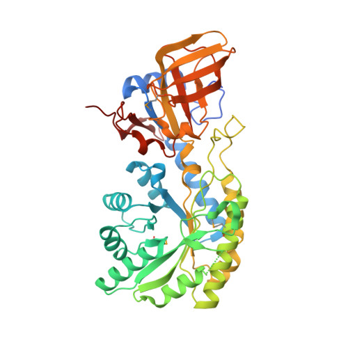
    The siderophore rhizoferrin (N 1 ,N 4 -dicitrylputrescine) is produced in fungi and bacteria to scavenge iron. Putrescine-producing bacterium Ralstonia pickettii synthesizes rhizoferrin and encodes a single nonribosomal peptide synthetase-independent siderophore (NIS) synthetase. From biosynthetic logic, we hypothesized that this single enzyme is sufficient for rhizoferrin biosynthesis. We confirmed this by expression of R. pickettii NIS synthetase in Escherichia coli, resulting in rhizoferrin production. This was further confirmed in vitro using the recombinant NIS synthetase, synthesizing rhizoferrin from putrescine and citrate. Heterologous expression of homologous lbtA from Legionella pneumophila, required for rhizoferrin biosynthesis in that species, produced siderophore activity in E. coli. Rhizoferrin is also synthesized by Francisella tularensis and Francisella novicida, but unlike R. pickettii or L. pneumophila, Francisella species lack putrescine biosynthetic pathways because of genomic decay. Francisella encodes a NIS synthetase FslA/FigA and an ornithine decarboxylase homolog FslC/FigC, required for rhizoferrin biosynthesis. Ornithine decarboxylase produces putrescine from ornithine, but we show here in vitro that FigA synthesizes N-citrylornithine, and FigC is an N-citrylornithine decarboxylase that together synthesize rhizoferrin without using putrescine. We co-expressed F. novicida figA and figC in E. coli and produced rhizoferrin. A 2.1 Å X-ray crystal structure of the FigC N-citrylornithine decarboxylase reveals how the larger substrate is accommodated and how active site residues have changed to recognize N-citrylornithine. FigC belongs to a new subfamily of alanine racemase-fold PLP-dependent decarboxylases that are not involved in polyamine biosynthesis. These data reveal a natural product biosynthetic workaround that evolved to bypass a missing precursor and re-establish it in the final structure.


  • Organizational Affiliation

    Department of Biochemistry, UT Southwestern Medical Center, Dallas, Texas, USA.


Macromolecules
Find similar proteins by:  (by identity cutoff)  |  3D Structure
Entity ID: 1
MoleculeChains Sequence LengthOrganismDetailsImage
N-citrylornithine decarboxylase
A, B, C, D
436Francisella tularensis subsp. novicidaMutation(s): 0 
Gene Names: figC
UniProt
Find proteins for Q15EG8 (Francisella novicida)
Explore Q15EG8 
Go to UniProtKB:  Q15EG8
Entity Groups  
Sequence Clusters30% Identity50% Identity70% Identity90% Identity95% Identity100% Identity
UniProt GroupQ15EG8
Sequence Annotations
Expand
  • Reference Sequence
Small Molecules
Ligands 3 Unique
IDChains Name / Formula / InChI Key2D Diagram3D Interactions
PLP (Subject of Investigation/LOI)
Query on PLP

Download Ideal Coordinates CCD File 
E [auth A],
H [auth B],
M [auth C],
P [auth D]
PYRIDOXAL-5'-PHOSPHATE
C8 H10 N O6 P
NGVDGCNFYWLIFO-UHFFFAOYSA-N
SO4
Query on SO4

Download Ideal Coordinates CCD File 
F [auth A]
G [auth A]
I [auth B]
J [auth B]
N [auth C]
F [auth A],
G [auth A],
I [auth B],
J [auth B],
N [auth C],
O [auth C],
Q [auth D],
R [auth D]
SULFATE ION
O4 S
QAOWNCQODCNURD-UHFFFAOYSA-L
GOL
Query on GOL

Download Ideal Coordinates CCD File 
K [auth B],
L [auth B]
GLYCEROL
C3 H8 O3
PEDCQBHIVMGVHV-UHFFFAOYSA-N
Modified Residues  1 Unique
IDChains TypeFormula2D DiagramParent
MSE
Query on MSE
A, B, C, D
L-PEPTIDE LINKINGC5 H11 N O2 SeMET
Experimental Data & Validation

Experimental Data

  • Method: X-RAY DIFFRACTION
  • Resolution: 2.05 Å
  • R-Value Free: 0.199 
  • R-Value Work: 0.174 
  • R-Value Observed: 0.174 
  • Space Group: P 21 21 2
Unit Cell:
Length ( Å )Angle ( ˚ )
a = 70.839α = 90
b = 279.061β = 90
c = 108.625γ = 90
Software Package:
Software NamePurpose
PHENIXrefinement
HKL-2000data reduction
HKL-2000data scaling
AMoREphasing

Structure Validation

View Full Validation Report


Ligand Structure Quality Assessment 


Entry History & Funding Information

Deposition Data


Funding OrganizationLocationGrant Number
National Institutes of Health/National Institute Of Allergy and Infectious Diseases (NIH/NIAID)United StatesR37AI034432

Revision History  (Full details and data files)

  • Version 1.0: 2020-12-16
    Type: Initial release
  • Version 1.1: 2021-06-30
    Changes: Database references
  • Version 1.2: 2021-07-28
    Changes: Database references