


Crystal Structure of Glucosamine-1-phosphate N-acetyltransferase from Stenotrophomonas maltophilia K279a
Abendroth, J., Lorimer, D.D., Horanyi, P.S., Edwards, T.E.To be published.
Experimental Data Snapshot
wwPDB Validation 3D Report Full Report

Entity ID: 1 | |||||
|---|---|---|---|---|---|
| Molecule | Chains | Sequence Length | Organism | Details | Image |
| Bifunctional protein GlmU | 463 | Stenotrophomonas maltophilia K279a | Mutation(s): 0 Gene Names: glmU, Smlt4108 EC: 2.7.7.23 (PDB Primary Data), 2.3.1.157 (PDB Primary Data) | 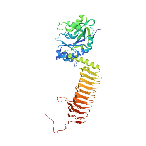 | |
UniProt | |||||
Find proteins for B2FHY5 (Stenotrophomonas maltophilia (strain K279a)) Explore B2FHY5 Go to UniProtKB: B2FHY5 | |||||
Entity Groups | |||||
| Sequence Clusters | 30% Identity50% Identity70% Identity90% Identity95% Identity100% Identity | ||||
| UniProt Group | B2FHY5 | ||||
Sequence AnnotationsExpand | |||||
| |||||
| Length ( Å ) | Angle ( ˚ ) |
|---|---|
| a = 91.29 | α = 90 |
| b = 91.29 | β = 90 |
| c = 184.55 | γ = 120 |
| Software Name | Purpose |
|---|---|
| XDS | data reduction |
| XSCALE | data scaling |
| PHENIX | refinement |
| PDB_EXTRACT | data extraction |
| MoRDa | phasing |
| PHENIX | model building |
| Coot | model building |
RCSB PDB (citation) is hosted by
RCSB PDB is a member of the

