7JQE

Structure of an extracellular fragment of EsaA from Streptococcus gallolyticus


Experimental Data Snapshot

  • Method: X-RAY DIFFRACTION
  • Resolution: 2.40 Å
  • R-Value Free: 0.262 
  • R-Value Work: 0.215 
  • R-Value Observed: 0.217 

wwPDB Validation   3D Report Full Report


This is version 1.2 of the entry. See complete history


Literature

Structure of the Extracellular Region of the Bacterial Type VIIb Secretion System Subunit EsaA.

Klein, T.A.Grebenc, D.W.Gandhi, S.Y.Shah, V.S.Kim, Y.Whitney, J.C.

(2021) Structure 29: 177-185.e6

  • DOI: https://doi.org/10.1016/j.str.2020.11.002
  • Primary Citation of Related Structures:  
    7JQE

  • PubMed Abstract: 

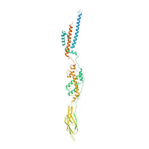
    Gram-positive bacteria use type VII secretion systems (T7SSs) to export effector proteins that manipulate the physiology of nearby prokaryotic and eukaryotic cells. Several mycobacterial T7SSs have established roles in virulence. By contrast, the genetically distinct T7SSb pathway found in Firmicutes bacteria more often functions to mediate bacterial competition. A lack of structural information on the T7SSb has limited the understanding of effector export by this protein secretion apparatus. Here, we present the 2.4 Å crystal structure of the extracellular region of the T7SSb subunit EsaA from Streptococcus gallolyticus. Our structure reveals that homodimeric EsaA is an elongated, arrow-shaped protein with a surface-accessible "tip", which in some species of bacteria serves as a receptor for lytic bacteriophages. Because it is the only T7SSb subunit large enough to traverse the peptidoglycan layer of Firmicutes, we propose that EsaA plays a critical role in transporting effectors across the entirety of the Gram-positive cell envelope.


  • Organizational Affiliation

    Michael DeGroote Institute for Infectious Disease Research, McMaster University, Hamilton, ON L8S 4K1, Canada; Department of Biochemistry and Biomedical Sciences, McMaster University, Hamilton, ON L8S 4K1, Canada.


Macromolecules
Find similar proteins by:  (by identity cutoff)  |  3D Structure
Entity ID: 1
MoleculeChains Sequence LengthOrganismDetailsImage
ESAT-6/WXG100 secretion system protein604Streptococcus gallolyticus subsp. gallolyticus ATCC 43143Mutation(s): 0 
Gene Names: esaASGGB_0524
Entity Groups  
Sequence Clusters30% Identity50% Identity70% Identity90% Identity95% Identity100% Identity
Sequence Annotations
Expand
  • Reference Sequence
Small Molecules
Ligands 2 Unique
IDChains Name / Formula / InChI Key2D Diagram3D Interactions
EDO
Query on EDO

Download Ideal Coordinates CCD File 
C [auth A]
D [auth A]
E [auth A]
F [auth A]
G [auth A]
C [auth A],
D [auth A],
E [auth A],
F [auth A],
G [auth A],
H [auth A],
I [auth A],
J [auth A],
K [auth A],
L [auth A],
M [auth A],
N [auth A],
O [auth A],
P [auth A],
Q [auth A],
R [auth A],
S [auth A],
T [auth A],
U [auth A],
V [auth A],
W [auth A],
X [auth A],
Y [auth A]
1,2-ETHANEDIOL
C2 H6 O2
LYCAIKOWRPUZTN-UHFFFAOYSA-N
CL
Query on CL

Download Ideal Coordinates CCD File 
B [auth A]CHLORIDE ION
Cl
VEXZGXHMUGYJMC-UHFFFAOYSA-M
Modified Residues  1 Unique
IDChains TypeFormula2D DiagramParent
MSE
Query on MSE
A
L-PEPTIDE LINKINGC5 H11 N O2 SeMET
Experimental Data & Validation

Experimental Data

  • Method: X-RAY DIFFRACTION
  • Resolution: 2.40 Å
  • R-Value Free: 0.262 
  • R-Value Work: 0.215 
  • R-Value Observed: 0.217 
  • Space Group: C 2 2 21
Unit Cell:
Length ( Å )Angle ( ˚ )
a = 74.466α = 90
b = 248.235β = 90
c = 81.095γ = 90
Software Package:
Software NamePurpose
PHENIXrefinement
DIALSdata reduction
DIALSdata scaling
HKL-3000phasing
PHENIXphasing
BUCCANEERmodel building

Structure Validation

View Full Validation Report


Entry History 

Deposition Data

Revision History  (Full details and data files)

  • Version 1.0: 2020-11-11
    Type: Initial release
  • Version 1.1: 2020-12-09
    Changes: Database references
  • Version 1.2: 2021-02-17
    Changes: Database references