7JLU

Structure of the activated Roq1 resistosome directly recognizing the pathogen effector XopQ


Experimental Data Snapshot

  • Method: ELECTRON MICROSCOPY
  • Resolution: 3.80 Å
  • Aggregation State: PARTICLE 
  • Reconstruction Method: SINGLE PARTICLE 

wwPDB Validation   3D Report Full Report


This is version 1.2 of the entry. See complete history


Literature

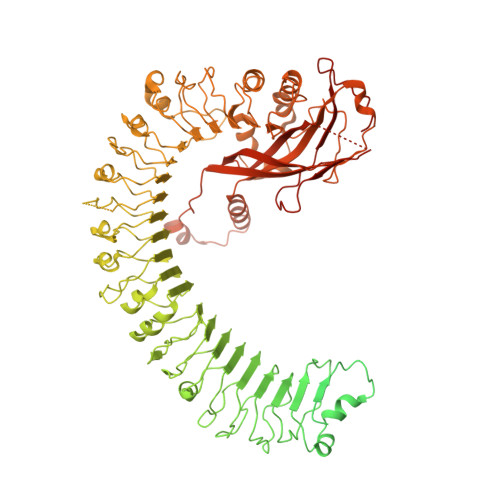
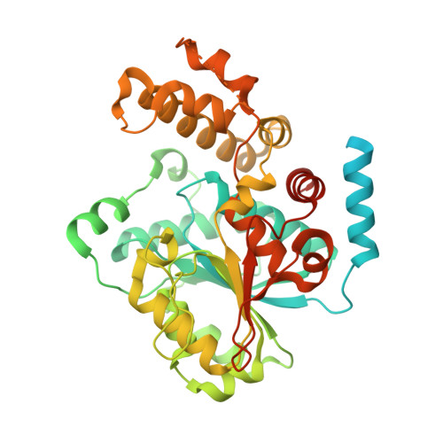
Structure of the activated ROQ1 resistosome directly recognizing the pathogen effector XopQ.

Martin, R.Qi, T.Zhang, H.Liu, F.King, M.Toth, C.Nogales, E.Staskawicz, B.J.

(2020) Science 370

  • DOI: https://doi.org/10.1126/science.abd9993
  • Primary Citation of Related Structures:  
    7JLU, 7JLV, 7JLX

  • PubMed Abstract: 

    Plants and animals detect pathogen infection using intracellular nucleotide-binding leucine-rich repeat receptors (NLRs) that directly or indirectly recognize pathogen effectors and activate an immune response. How effector sensing triggers NLR activation remains poorly understood. Here we describe the 3.8-angstrom-resolution cryo-electron microscopy structure of the activated ROQ1 (recognition of XopQ 1), an NLR native to Nicotiana benthamiana with a Toll-like interleukin-1 receptor (TIR) domain bound to the Xanthomonas euvesicatoria effector XopQ ( Xanthomonas outer protein Q). ROQ1 directly binds to both the predicted active site and surface residues of XopQ while forming a tetrameric resistosome that brings together the TIR domains for downstream immune signaling. Our results suggest a mechanism for the direct recognition of effectors by NLRs leading to the oligomerization-dependent activation of a plant resistosome and signaling by the TIR domain.


  • Organizational Affiliation

    Biophysics Graduate Group, University of California, Berkeley, CA 94720, USA.


Macromolecules
Find similar proteins by:  (by identity cutoff)  |  3D Structure
Entity ID: 1
MoleculeChains Sequence LengthOrganismDetailsImage
Disease resistance protein Roq11,328Nicotiana benthamianaMutation(s): 0 
Gene Names: ROQ1
EC: 3.2.2.6
UniProt
Find proteins for A0A290U7C4 (Nicotiana benthamiana)
Explore A0A290U7C4 
Go to UniProtKB:  A0A290U7C4
Entity Groups  
Sequence Clusters30% Identity50% Identity70% Identity90% Identity95% Identity100% Identity
UniProt GroupA0A290U7C4
Sequence Annotations
Expand
  • Reference Sequence
Find similar proteins by:  (by identity cutoff)  |  3D Structure
Entity ID: 2
MoleculeChains Sequence LengthOrganismDetailsImage
XopQ477Xanthomonas euvesicatoriaMutation(s): 0 
Gene Names: xopQ
UniProt
Find proteins for Q5QA88 (Xanthomonas euvesicatoria)
Explore Q5QA88 
Go to UniProtKB:  Q5QA88
Entity Groups  
Sequence Clusters30% Identity50% Identity70% Identity90% Identity95% Identity100% Identity
UniProt GroupQ5QA88
Sequence Annotations
Expand
  • Reference Sequence
Small Molecules
Ligands 1 Unique
IDChains Name / Formula / InChI Key2D Diagram3D Interactions
CA (Subject of Investigation/LOI)
Query on CA

Download Ideal Coordinates CCD File 
C [auth B]CALCIUM ION
Ca
BHPQYMZQTOCNFJ-UHFFFAOYSA-N
Experimental Data & Validation

Experimental Data

  • Method: ELECTRON MICROSCOPY
  • Resolution: 3.80 Å
  • Aggregation State: PARTICLE 
  • Reconstruction Method: SINGLE PARTICLE 
EM Software:
TaskSoftware PackageVersion
RECONSTRUCTIONRELION3.1
MODEL REFINEMENTPHENIX1.18

Structure Validation

View Full Validation Report


Entry History & Funding Information

Deposition Data


Funding OrganizationLocationGrant Number
Howard Hughes Medical Institute (HHMI)United States--

Revision History  (Full details and data files)

  • Version 1.0: 2020-12-02
    Type: Initial release
  • Version 1.1: 2020-12-16
    Changes: Database references
  • Version 1.2: 2024-03-06
    Changes: Data collection, Database references