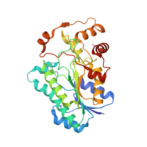
7JID

Crystal structure of the L780 UDP-rhamnose synthase from Acanthamoeba polyphaga mimivirus


Experimental Data Snapshot

  • Method: X-RAY DIFFRACTION
  • Resolution: 1.45 Å
  • R-Value Free: 0.227 
  • R-Value Work: 0.198 
  • R-Value Observed: 0.199 

wwPDB Validation   3D Report Full Report


Ligand Structure Quality Assessment 


This is version 1.2 of the entry. See complete history


Literature

The high-resolution structure of a UDP-L-rhamnose synthase from Acanthamoeba polyphaga Mimivirus.

Bockhaus, N.J.Ferek, J.D.Thoden, J.B.Holden, H.M.

(2020) Protein Sci 29: 2164-2174

  • DOI: https://doi.org/10.1002/pro.3928
  • Primary Citation of Related Structures:  
    7JID

  • PubMed Abstract: 

    For the field of virology, perhaps one of the most paradigm-shifting events so far in the 21st century was the identification of the giant double-stranded DNA virus that infects amoebae. Remarkably, this virus, known as Mimivirus, has a genome that encodes for nearly 1,000 proteins, some of which are involved in the biosynthesis of unusual sugars. Indeed, the virus is coated by a layer of glycosylated fibers that contain d-glucose, N-acetyl-d-glucosamine, l-rhamnose, and 4-amino-4,6-dideoxy-d-glucose. Here we describe a combined structural and enzymological investigation of the protein encoded by the open-reading frame L780, which corresponds to an l-rhamnose synthase. The structure of the L780/NADP + /UDP-l-rhamnose ternary complex was determined to 1.45 Å resolution and refined to an overall R-factor of 19.9%. Each subunit of the dimeric protein adopts a bilobal-shaped appearance with the N-terminal domain harboring the dinucleotide-binding site and the C-terminal domain positioning the UDP-sugar into the active site. The overall molecular architecture of L780 places it into the short-chain dehydrogenase/reductase superfamily. Kinetic analyses indicate that the enzyme can function on either UDP- and dTDP-sugars but displays a higher catalytic efficiency with the UDP-linked substrate. Site-directed mutagenesis experiments suggest that both Cys 108 and Lys 175 play key roles in catalysis. This structure represents the first model of a viral UDP-l-rhamnose synthase and provides new details into these fascinating enzymes.


  • Organizational Affiliation

    Department of Biochemistry, University of Wisconsin, Madison, Wisconsin, USA.


Macromolecules
Find similar proteins by:  (by identity cutoff)  |  3D Structure
Entity ID: 1
MoleculeChains Sequence LengthOrganismDetailsImage
UDP-L-Rhamnose Synthase
A, B
292Acanthamoeba polyphaga mimivirusMutation(s): 0 
Gene Names: MIMI_L780
UniProt
Find proteins for Q5UPS5 (Acanthamoeba polyphaga mimivirus)
Explore Q5UPS5 
Go to UniProtKB:  Q5UPS5
Entity Groups  
Sequence Clusters30% Identity50% Identity70% Identity90% Identity95% Identity100% Identity
UniProt GroupQ5UPS5
Sequence Annotations
Expand
  • Reference Sequence
Small Molecules
Ligands 3 Unique
IDChains Name / Formula / InChI Key2D Diagram3D Interactions
NAP
Query on NAP

Download Ideal Coordinates CCD File 
D [auth A],
L [auth B]
NADP NICOTINAMIDE-ADENINE-DINUCLEOTIDE PHOSPHATE
C21 H28 N7 O17 P3
XJLXINKUBYWONI-NNYOXOHSSA-N
AWU (Subject of Investigation/LOI)
Query on AWU

Download Ideal Coordinates CCD File 
C [auth A],
K [auth B]
[[(2~{R},3~{S},4~{R},5~{R})-5-[2,4-bis(oxidanylidene)pyrimidin-1-yl]-3,4-bis(oxidanyl)oxolan-2-yl]methoxy-oxidanyl-phosphoryl] [(2~{R},3~{R},4~{R},5~{R},6~{S})-6-methyl-3,4,5-tris(oxidanyl)oxan-2-yl] hydrogen phosphate
C15 H24 N2 O16 P2
DRDCJEIZVLVWNC-SLBWPEPYSA-N
EDO
Query on EDO

Download Ideal Coordinates CCD File 
E [auth A]
F [auth A]
G [auth A]
H [auth A]
I [auth A]
E [auth A],
F [auth A],
G [auth A],
H [auth A],
I [auth A],
J [auth A],
M [auth B],
N [auth B],
O [auth B]
1,2-ETHANEDIOL
C2 H6 O2
LYCAIKOWRPUZTN-UHFFFAOYSA-N
Experimental Data & Validation

Experimental Data

  • Method: X-RAY DIFFRACTION
  • Resolution: 1.45 Å
  • R-Value Free: 0.227 
  • R-Value Work: 0.198 
  • R-Value Observed: 0.199 
  • Space Group: P 1
Unit Cell:
Length ( Å )Angle ( ˚ )
a = 49.803α = 81.94
b = 52.989β = 88.68
c = 70.149γ = 66.74
Software Package:
Software NamePurpose
REFMACrefinement
PDB_EXTRACTdata extraction
SAINTdata reduction
SADABSdata scaling
PHASERphasing

Structure Validation

View Full Validation Report


Ligand Structure Quality Assessment 


Entry History & Funding Information

Deposition Data


Funding OrganizationLocationGrant Number
National Institutes of Health/National Institute of General Medical Sciences (NIH/NIGMS)United StatesGM134643

Revision History  (Full details and data files)

  • Version 1.0: 2020-08-26
    Type: Initial release
  • Version 1.1: 2020-11-04
    Changes: Database references
  • Version 1.2: 2023-10-18
    Changes: Data collection, Database references, Refinement description