Insights into Two-Metal-Ion Catalytic Mechanism of Cap-Snatching Endonuclease of Ebinur Lake Virus in Bunyavirales.
Kuang, W., Zhang, H., Cai, Y., Zhang, G., Deng, F., Li, H., Hu, Z., Guo, Y., Wang, M., Zhou, Y., Gong, P.(2022) J Virol 96: e0208521-e0208521
- PubMed: 35044209
- DOI: https://doi.org/10.1128/jvi.02085-21
- Primary Citation of Related Structures:
7EWU, 7EWV, 7EWW, 7EWX, 7EWY, 7EWZ, 7EX0, 7EX1 - PubMed Abstract:
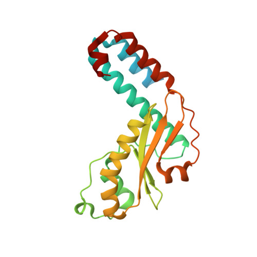
The cap-snatching endonuclease (EN) of segmented negative-strand RNA viruses (sNSVs) produces short capped primers for viral transcription by cleaving the host mRNAs. EN requires divalent metals as cofactors for nucleic acid substrates cleavage; however, the detailed mechanism of metal ion-dependent catalysis of ENs remains obscure. In this work, we reported the EN crystal structure of the Ebinur Lake virus (EBIV), an emerging mosquito-borne orthobunyavirus, and investigated its enzymatic properties and metal ion-based catalytic mechanism. In vitro biochemical data showed that EBIV EN is a specific RNA nuclease and prefers to cleave unstructured uridine-rich ssRNA. Structural comparison indicated that the overall structural architecture of EBIV EN is similar to that of other sNSV ENs, while the detailed active site configuration including the binding state of metal ions and the conformation of the LA/LB loop pair is different. Based on sequence conservation analysis, nine active site mutants were constructed, and seven crystal structures of them were determined. Mutations of active site residues associated with the two metal ions (Mn1 and Mn2) coordination abolished EN activity. Crystallographic analyses further revealed that none of these mutants bound two metal ions simultaneously in the active site. Importantly, we found that the perturbation of Mn1-coordination (metal site 1), resulted in the enhancement or elimination of Mn2-coordination (metal site 2). Taken together, our data provide structural evidence to support the two-metal-ion catalytic mechanism of EBIV EN and the correlation of metal binding at the two binding sites, which may be commonly shared by bunyaviruses or other sNSVs. IMPORTANCE The viral endonucleases (ENs) encoded by bunyaviruses and orthomyxoviruses play an essential role in initiating transcription by "snatching" capped primers from the host mRNAs. These ENs are metal-ion-dependent nucleases; however, the details of their catalytic mechanism remain elusive. Here, we reported high-resolution crystal structures of the wild-type and mutant ENs of a novel bunyavirus, the Ebinur Lake virus (EBIV), and revealed the structure and function relationship of EN. The EBIV EN exhibited differences in the details of active site structure compared to its homologues. Our data provided structural evidence to support a two-metal-ion catalytic mechanism of EBIV EN, and found the correlation of metal binding at both binding sites, which might reflect the dynamic structural properties that correlate to EN catalytic function. Taken together, our results revealed the structural characteristics of EBIV EN and made important implications for understanding the catalytic mechanism of cap-snatching ENs.
Organizational Affiliation:
Department of Forensic Medicine, Tongji Medical College, Huazhong University of Science and Technology, Wuhan, Hubei, China.