7CV2

Crystal structure of B. halodurans NiaR in niacin-bound form


Experimental Data Snapshot

  • Method: X-RAY DIFFRACTION
  • Resolution: 1.80 Å
  • R-Value Free: 0.243 
  • R-Value Work: 0.194 
  • R-Value Observed: 0.197 

wwPDB Validation   3D Report Full Report


Ligand Structure Quality Assessment 


This is version 1.1 of the entry. See complete history


Literature

Structural analysis and insight into effector binding of the niacin-responsive repressor NiaR from Bacillus halodurans.

Lee, D.W.Park, Y.W.Lee, M.Y.Jeong, K.H.Lee, J.Y.

(2020) Sci Rep 10: 21039-21039

  • DOI: https://doi.org/10.1038/s41598-020-78148-x
  • Primary Citation of Related Structures:  
    7CV0, 7CV2

  • PubMed Abstract: 

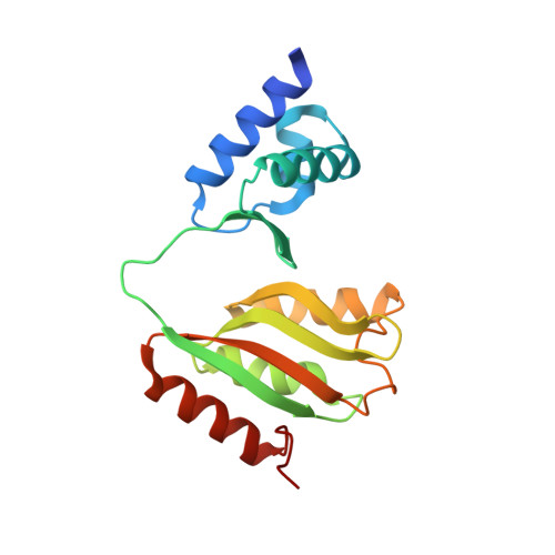
    The niacin-responsive repressor, NiaR, is transcriptional repressor of certain nicotinamide adenine dinucleotide (NAD) biosynthetic genes in response to an increase in niacin levels. NAD is a vital molecule involved in various cellular redox reactions as an electron donor or electron acceptor. The NiaR family is conserved broadly in the Bacillus/Clostridium group, as well as in the Fusobacteria and Thermotogales lineages. The NiaR structure consists of two domains: an N-terminal DNA-binding domain, and a C-terminal regulation domain containing a metal-binding site. In this paper, we report the crystal structures of apo and niacin-bound forms of NiaR from Bacillus halodurans (BhNiaR). The analysis of metal-binding and niacin-binding sites through the apo and niacin-bound structures is described. Each N- and C-terminal domain structure of BhNiaR is almost identical with NiaR from Thermotoga maritima, but the overall domain arrangement is quite different. A zinc ion is fully occupied in each subunit with well-conserved residues in the C-terminal domain. Niacin is also located at a hydrophobic pocket near the zinc ion in the C-terminal domain.


  • Organizational Affiliation

    Department of Life Science, Dongguk University-Seoul, Ilsandong-gu, Goyang-si, Gyeonggi-do, 10326, Republic of Korea.


Macromolecules
Find similar proteins by:  (by identity cutoff)  |  3D Structure
Entity ID: 1
MoleculeChains Sequence LengthOrganismDetailsImage
Transcriptional regulator NiaR179Halalkalibacterium halodurans C-125Mutation(s): 0 
Gene Names: BH1216
UniProt
Find proteins for Q9KDJ7 (Halalkalibacterium halodurans (strain ATCC BAA-125 / DSM 18197 / FERM 7344 / JCM 9153 / C-125))
Explore Q9KDJ7 
Go to UniProtKB:  Q9KDJ7
Entity Groups  
Sequence Clusters30% Identity50% Identity70% Identity90% Identity95% Identity100% Identity
UniProt GroupQ9KDJ7
Sequence Annotations
Expand
  • Reference Sequence
Experimental Data & Validation

Experimental Data

  • Method: X-RAY DIFFRACTION
  • Resolution: 1.80 Å
  • R-Value Free: 0.243 
  • R-Value Work: 0.194 
  • R-Value Observed: 0.197 
  • Space Group: P 43 21 2
Unit Cell:
Length ( Å )Angle ( ˚ )
a = 42.249α = 90
b = 42.249β = 90
c = 175.99γ = 90
Software Package:
Software NamePurpose
PHENIXrefinement
HKL-2000data reduction
HKL-2000data scaling
PHASERphasing

Structure Validation

View Full Validation Report


Ligand Structure Quality Assessment 


Entry History 

Deposition Data

Revision History  (Full details and data files)

  • Version 1.0: 2020-12-16
    Type: Initial release
  • Version 1.1: 2023-11-29
    Changes: Data collection, Database references, Refinement description