7CNA

Crystal structure of Spindlin1/C11orf84 complex bound to histone H3K4me3K9me3 peptide


Experimental Data Snapshot

  • Method: X-RAY DIFFRACTION
  • Resolution: 1.60 Å
  • R-Value Free: 0.201 
  • R-Value Work: 0.172 
  • R-Value Observed: 0.173 

wwPDB Validation   3D Report Full Report


This is version 2.1 of the entry. See complete history


Literature

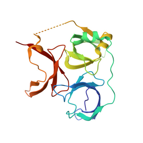
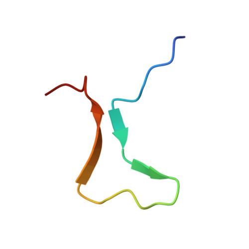
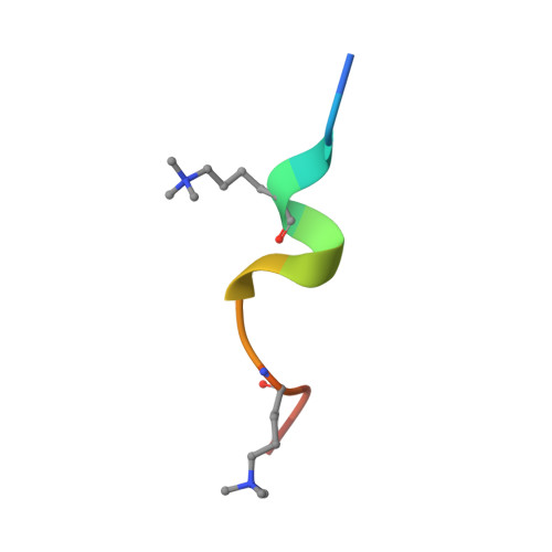
Structural mechanism of bivalent histone H3K4me3K9me3 recognition by the Spindlin1/C11orf84 complex in rRNA transcription activation.

Du, Y.Yan, Y.Xie, S.Huang, H.Wang, X.Ng, R.K.Zhou, M.M.Qian, C.

(2021) Nat Commun 12: 949-949

  • DOI: https://doi.org/10.1038/s41467-021-21236-x
  • Primary Citation of Related Structures:  
    7CNA

  • PubMed Abstract: 

    Spindlin1 is a unique multivalent epigenetic reader that facilitates ribosomal RNA transcription. In this study, we provide molecular and structural basis by which Spindlin1 acts in complex with C11orf84 to preferentially recognize non-canonical bivalent mark of trimethylated lysine 4 and lysine 9 present on the same histone H3 tail (H3K4me3K9me3). We demonstrate that C11orf84 binding stabilizes Spindlin1 and enhances its association with bivalent H3K4me3K9me3 mark. The functional analysis suggests that Spindlin1/C11orf84 complex can displace HP1 proteins from H3K4me3K9me3-enriched rDNA loci, thereby facilitating the conversion of these poised rDNA repeats from the repressed state to the active conformation, and the consequent recruitment of RNA Polymerase I for rRNA transcription. Our study uncovers a previously unappreciated mechanism of bivalent H3K4me3K9me3 recognition by Spindlin1/C11orf84 complex required for activation of rRNA transcription.


  • Organizational Affiliation

    School of Biomedical Sciences, The University of Hong Kong, Hong Kong Island, Hong Kong.


Macromolecules
Find similar proteins by:  (by identity cutoff)  |  3D Structure
Entity ID: 1
MoleculeChains Sequence LengthOrganismDetailsImage
Spindlin-1A,
C [auth D]
212Homo sapiensMutation(s): 0 
Gene Names: SPIN1OCRSPIN
UniProt & NIH Common Fund Data Resources
Find proteins for Q9Y657 (Homo sapiens)
Explore Q9Y657 
Go to UniProtKB:  Q9Y657
PHAROS:  Q9Y657
GTEx:  ENSG00000106723 
Entity Groups  
Sequence Clusters30% Identity50% Identity70% Identity90% Identity95% Identity100% Identity
UniProt GroupQ9Y657
Sequence Annotations
Expand
  • Reference Sequence
Find similar proteins by:  (by identity cutoff)  |  3D Structure
Entity ID: 2
MoleculeChains Sequence LengthOrganismDetailsImage
Spindlin interactor and repressor of chromatin-binding proteinB,
D [auth E]
30Homo sapiensMutation(s): 0 
Gene Names: SPINDOCC11orf84
UniProt & NIH Common Fund Data Resources
Find proteins for Q9BUA3 (Homo sapiens)
Explore Q9BUA3 
Go to UniProtKB:  Q9BUA3
PHAROS:  Q9BUA3
GTEx:  ENSG00000168005 
Entity Groups  
Sequence Clusters30% Identity50% Identity70% Identity90% Identity95% Identity100% Identity
UniProt GroupQ9BUA3
Sequence Annotations
Expand
  • Reference Sequence

Find similar proteins by:  Sequence   |   3D Structure  

Entity ID: 3
MoleculeChains Sequence LengthOrganismDetailsImage
ALA-ARG-THR-M3L-GLN-THR-ALA-ARG-M3L-SER-THRE [auth C]12synthetic constructMutation(s): 0 
Entity Groups  
Sequence Clusters30% Identity50% Identity70% Identity90% Identity95% Identity100% Identity
Sequence Annotations
Expand
  • Reference Sequence

Find similar proteins by:  Sequence   |   3D Structure  

Entity ID: 4
MoleculeChains Sequence LengthOrganismDetailsImage
ALA-ARG-THR-M3L-GLN-THR-ALA-ARG-M3L-SER-GLY12synthetic constructMutation(s): 0 
Entity Groups  
Sequence Clusters30% Identity50% Identity70% Identity90% Identity95% Identity100% Identity
Sequence Annotations
Expand
  • Reference Sequence
Small Molecules
Ligands 3 Unique
IDChains Name / Formula / InChI Key2D Diagram3D Interactions
BEN
Query on BEN

Download Ideal Coordinates CCD File 
G [auth A]
H [auth A]
J [auth D]
K [auth D]
L [auth D]
G [auth A],
H [auth A],
J [auth D],
K [auth D],
L [auth D],
P [auth C]
BENZAMIDINE
C7 H8 N2
PXXJHWLDUBFPOL-UHFFFAOYSA-N
GOL
Query on GOL

Download Ideal Coordinates CCD File 
N [auth E]GLYCEROL
C3 H8 O3
PEDCQBHIVMGVHV-UHFFFAOYSA-N
CL
Query on CL

Download Ideal Coordinates CCD File 
I [auth A],
M [auth D],
O [auth E]
CHLORIDE ION
Cl
VEXZGXHMUGYJMC-UHFFFAOYSA-M
Modified Residues  1 Unique
IDChains TypeFormula2D DiagramParent
M3L
Query on M3L
E [auth C]L-PEPTIDE LINKINGC9 H21 N2 O2LYS
Experimental Data & Validation

Experimental Data

  • Method: X-RAY DIFFRACTION
  • Resolution: 1.60 Å
  • R-Value Free: 0.201 
  • R-Value Work: 0.172 
  • R-Value Observed: 0.173 
  • Space Group: P 1 21 1
Unit Cell:
Length ( Å )Angle ( ˚ )
a = 45.603α = 90
b = 101.184β = 91.41
c = 56.615γ = 90
Software Package:
Software NamePurpose
REFMACrefinement
XSCALEdata scaling
PHASERphasing
PDB_EXTRACTdata extraction
XDSdata reduction

Structure Validation

View Full Validation Report


Entry History & Funding Information

Deposition Data

  • Released Date: 2021-01-13 
  • Deposition Author(s): Qian, C.M.

Funding OrganizationLocationGrant Number
The University Grants Committee, Research Grants Council (RGC)Hong Kong17127917

Revision History  (Full details and data files)

  • Version 1.0: 2021-01-13
    Type: Initial release
  • Version 1.1: 2021-08-11
    Changes: Database references
  • Version 2.0: 2021-08-18
    Changes: Atomic model, Data collection, Derived calculations, Non-polymer description, Structure summary
  • Version 2.1: 2023-11-29
    Changes: Data collection, Refinement description