7CFL

X-ray structure of autolysin Acd24020 catalytic domain from Clostridium difficile


Experimental Data Snapshot

  • Method: X-RAY DIFFRACTION
  • Resolution: 1.56 Å
  • R-Value Free: 0.258 
  • R-Value Work: 0.198 
  • R-Value Observed: 0.201 

wwPDB Validation   3D Report Full Report


Ligand Structure Quality Assessment 


This is version 1.2 of the entry. See complete history


Literature

Structural and biochemical characterizations of the novel autolysin Acd24020 from Clostridioides difficile and its full-function catalytic domain as a lytic enzyme.

Sekiya, H.Tamai, E.Kawasaki, J.Murakami, K.Kamitori, S.

(2021) Mol Microbiol 115: 684-698

  • DOI: https://doi.org/10.1111/mmi.14636
  • Primary Citation of Related Structures:  
    7CFL

  • PubMed Abstract: 

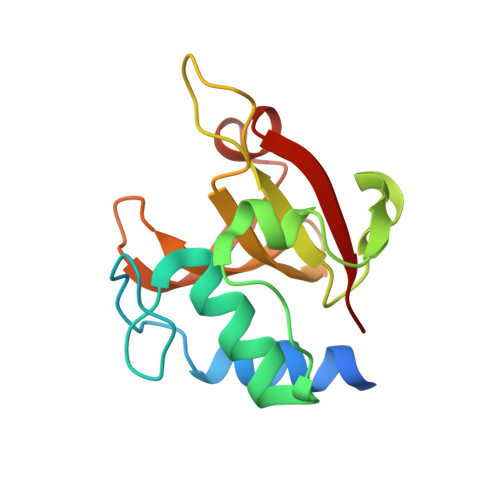
    Autolysin is a lytic enzyme that hydrolyzes peptidoglycans of the bacterial cell wall, with a catalytic domain and cell wall-binding (CWB) domains, to be involved in different physiological functions that require bacterial cell wall remodeling. We identified a novel autolysin, Acd24020, from Clostridioides (Clostridium) difficile (C. difficile), with an endopeptidase catalytic domain belonging to the NlpC/P60 family and three bacterial Src-homology 3 domains as CWB domains. The catalytic domain of Acd24020 (Acd24020-CD) exhibited C. difficile-specific lytic activity equivalent to Acd24020, indicating that Acd24020-CD has full-function as a lytic enzyme by itself. To elucidate the specific peptidoglycan-recognition and catalytic reaction mechanisms of Acd24020-CD, biochemical characterization, X-ray structure determination, a modeling study of the enzyme/substrate complex, and mutagenesis analysis were performed. Acd24020-CD has an hourglass-shaped substrate-binding groove across the molecule, which is responsible for recognizing the direct 3-4 cross-linking structure unique to C. difficile peptidoglycan. Based on the X-ray structure and modeling study, we propose a dynamic Cys/His catalyzing mechanism, in which the catalytic Cys299 and His354 residues dynamically change their conformations to complement each step of the enzyme catalytic reaction.


  • Organizational Affiliation

    Department of Infectious Disease, College of Pharmaceutical Science, Matsuyama University, Matsuyama, Japan.


Macromolecules
Find similar proteins by:  (by identity cutoff)  |  3D Structure
Entity ID: 1
MoleculeChains Sequence LengthOrganismDetailsImage
Putative cell wall hydrolase phosphatase-associated protein
A, B, C, D
138Clostridioides difficileMutation(s): 0 
Gene Names: CD630_24020
EC: 3.4
UniProt
Find proteins for Q181Y8 (Clostridioides difficile (strain 630))
Explore Q181Y8 
Go to UniProtKB:  Q181Y8
Entity Groups  
Sequence Clusters30% Identity50% Identity70% Identity90% Identity95% Identity100% Identity
UniProt GroupQ181Y8
Sequence Annotations
Expand
  • Reference Sequence
Experimental Data & Validation

Experimental Data

  • Method: X-RAY DIFFRACTION
  • Resolution: 1.56 Å
  • R-Value Free: 0.258 
  • R-Value Work: 0.198 
  • R-Value Observed: 0.201 
  • Space Group: P 1
Unit Cell:
Length ( Å )Angle ( ˚ )
a = 50.44α = 104.86
b = 55.21β = 112.64
c = 57.11γ = 90.06
Software Package:
Software NamePurpose
REFMACrefinement
XDSdata reduction
XDSdata scaling
MOLREPphasing

Structure Validation

View Full Validation Report


Ligand Structure Quality Assessment 


Entry History & Funding Information

Deposition Data


Funding OrganizationLocationGrant Number
Japan Society for the Promotion of Science (JSPS)Japan18K06087

Revision History  (Full details and data files)

  • Version 1.0: 2020-11-25
    Type: Initial release
  • Version 1.1: 2021-05-12
    Changes: Database references
  • Version 1.2: 2023-11-29
    Changes: Data collection, Database references, Refinement description