Crystal structure and DNA cleavage mechanism of the restriction DNA glycosylase R.CcoLI from Campylobacter coli.
Miyazono, K.I., Wang, D., Ito, T., Tanokura, M.(2021) Sci Rep 11: 859-859
- PubMed: 33441677
- DOI: https://doi.org/10.1038/s41598-020-79537-y
- Primary Citation of Related Structures:
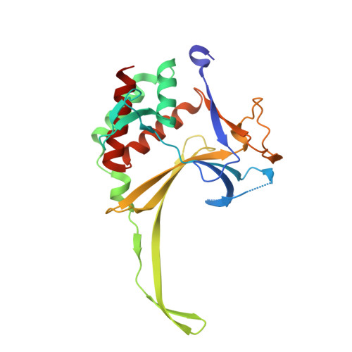
7CFA - PubMed Abstract:
While most restriction enzymes catalyze the hydrolysis of phosphodiester bonds at specific nucleotide sequences in DNA, restriction enzymes of the HALFPIPE superfamily cleave N-glycosidic bonds, similar to DNA glycosylases. Apurinic/apyrimidinic (AP) sites generated by HALFPIPE superfamily proteins are cleaved by their inherent AP lyase activities, other AP endonuclease activities or heat-promoted β-elimination. Although the HALFPIPE superfamily protein R.PabI, obtained from a hyperthermophilic archaea, Pyrococcus abyssi, shows weak AP lyase activity, HALFPIPE superfamily proteins in mesophiles, such as R.CcoLI from Campylobacter coli and R. HpyAXII from Helicobacter pylori, show significant AP lyase activities. To identify the structural basis for the AP lyase activity of R.CcoLI, we determined the structure of R.CcoLI by X-ray crystallography. The structure of R.CcoLI, obtained at 2.35-Å resolution, shows that a conserved lysine residue (Lys71), which is stabilized by a characteristic β-sheet structure of R.CcoLI, protrudes into the active site. The results of mutational assays indicate that Lys71 is important for the AP lyase activity of R.CcoLI. Our results help to elucidate the mechanism by which HALFPIPE superfamily proteins from mesophiles efficiently introduce double-strand breaks to specific sites on double-stranded DNA.
Organizational Affiliation:
Department of Applied Biological Chemistry, Graduate School of Agricultural and Life Sciences, The University of Tokyo, 1-1-1 Yayoi Bunkyo-ku, Tokyo, 113-8657, Japan.