7C6O

Catalytic Subunit of Cobaltochelatase from Mycobacterium tuberculosis

  • Classification: LIGASE
  • Organism(s): Mycobacterium tuberculosis
  • Expression System: Escherichia coli
  • Mutation(s): No 

  • Deposited: 2020-05-22 Released: 2020-12-02 
  • Deposition Author(s): Zhang, J., Liu, L.
  • Funding Organization(s): National Natural Science Foundation of China (NSFC), Ministry of Science and Technology (MoST, China)

Experimental Data Snapshot

  • Method: X-RAY DIFFRACTION
  • Resolution: 1.80 Å
  • R-Value Free: 0.196 
  • R-Value Work: 0.163 
  • R-Value Observed: 0.165 

wwPDB Validation   3D Report Full Report


This is version 1.2 of the entry. See complete history


Literature

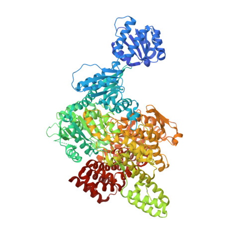
Crystal structure of the large subunit of cobaltochelatase from Mycobacterium tuberculosis.

Zhang, J.H.Yuan, H.Wang, X.Dai, H.E.Zhang, M.Liu, L.

(2021) Proteins 89: 462-467

  • DOI: https://doi.org/10.1002/prot.26023
  • Primary Citation of Related Structures:  
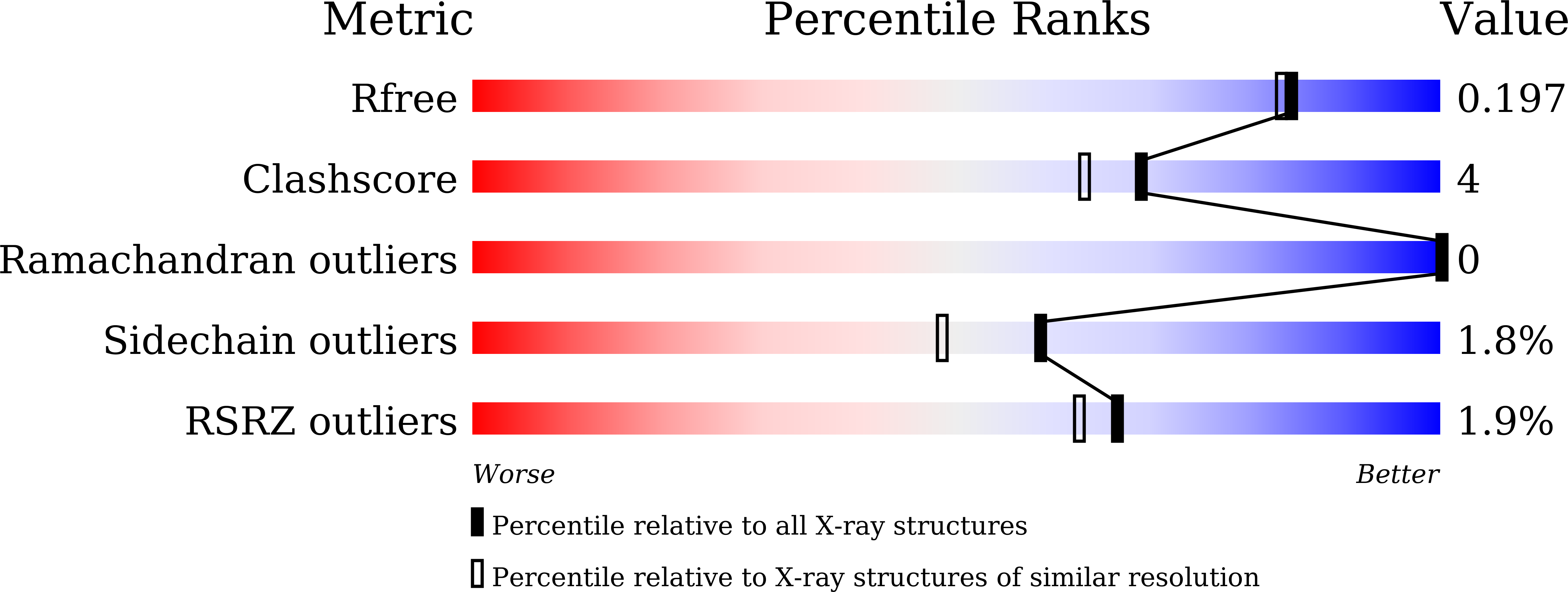
    7C6O

  • PubMed Abstract: 

    Cobaltochelatase in aerobic cobalamin biosynthesis is a complex composed of three subunits. The large subunit CobN is a 140-kDa protein and is homologous to the ChlH subunit of magnesium chelatase. Previously we have reported the 2.5-Å structure of a cyanobacterial ChlH. Here we present the 1.8-Å structure of CobN from Mycobacterium tuberculosis. The overall structure of CobN and ChlH is similar, but significant difference occurs in the head domain. Structural comparison of domains between the two proteins unravels candidate regions for substrate binding and helps to locate a triad of residues that may be essential for metal ion binding.


  • Organizational Affiliation

    School of Life Sciences, Anhui University, Hefei, China.


Macromolecules
Find similar proteins by:  (by identity cutoff)  |  3D Structure
Entity ID: 1
MoleculeChains Sequence LengthOrganismDetailsImage
Cobalamin biosynthesis protein CobN1,230Mycobacterium tuberculosisMutation(s): 0 
Gene Names: cobNDSI38_03330ERS023446_03562ERS094182_01916F6W99_00653GJE03_10820
EC: 6.6.1.2
UniProt
Find proteins for O53498 (Mycobacterium tuberculosis (strain ATCC 25618 / H37Rv))
Explore O53498 
Go to UniProtKB:  O53498
Entity Groups  
Sequence Clusters30% Identity50% Identity70% Identity90% Identity95% Identity100% Identity
UniProt GroupO53498
Sequence Annotations
Expand
  • Reference Sequence
Experimental Data & Validation

Experimental Data

  • Method: X-RAY DIFFRACTION
  • Resolution: 1.80 Å
  • R-Value Free: 0.196 
  • R-Value Work: 0.163 
  • R-Value Observed: 0.165 
  • Space Group: P 1 21 1
Unit Cell:
Length ( Å )Angle ( ˚ )
a = 65.564α = 90
b = 125.473β = 103.23
c = 78.551γ = 90
Software Package:
Software NamePurpose
PHENIXrefinement
HKL-2000data scaling
PDB_EXTRACTdata extraction
HKL-3000data reduction
HKL-3000phasing

Structure Validation

View Full Validation Report


Entry History & Funding Information

Deposition Data


Funding OrganizationLocationGrant Number
National Natural Science Foundation of China (NSFC)China31870729
Ministry of Science and Technology (MoST, China)China2017YFA0503703

Revision History  (Full details and data files)

  • Version 1.0: 2020-12-02
    Type: Initial release
  • Version 1.1: 2021-03-17
    Changes: Database references
  • Version 1.2: 2023-11-29
    Changes: Data collection, Database references, Refinement description