


Crystal structure of Serine acetyltransferase isoform 3 in complex with cysteine from Entamoeba histolytica
Dharavath, S., Kumar, S.To be published.
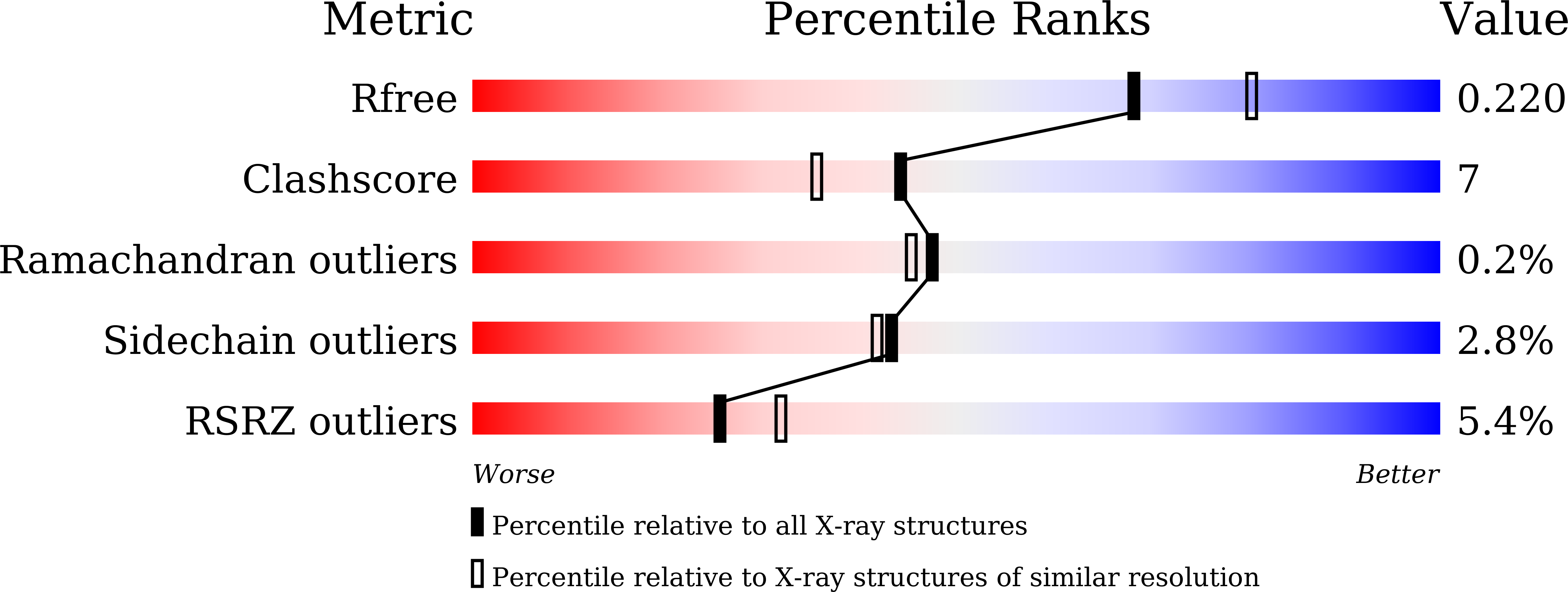
Experimental Data Snapshot
Entity ID: 1 | |||||
|---|---|---|---|---|---|
| Molecule | Chains | Sequence Length | Organism | Details | Image |
| Serine acetyltransferase 1, putative | 336 | Entamoeba histolytica | Mutation(s): 0 Gene Names: KM1_023830 |  | |
UniProt | |||||
Find proteins for Q401L4 (Entamoeba histolytica) Explore Q401L4 Go to UniProtKB: Q401L4 | |||||
Entity Groups | |||||
| Sequence Clusters | 30% Identity50% Identity70% Identity90% Identity95% Identity100% Identity | ||||
| UniProt Group | Q401L4 | ||||
Sequence AnnotationsExpand | |||||
| |||||
| Ligands 1 Unique | |||||
|---|---|---|---|---|---|
| ID | Chains | Name / Formula / InChI Key | 2D Diagram | 3D Interactions | |
| CYS (Subject of Investigation/LOI) Query on CYS | D [auth A], E [auth A], F [auth B] | CYSTEINE C3 H7 N O2 S XUJNEKJLAYXESH-REOHCLBHSA-N |  | ||
| Length ( Å ) | Angle ( ˚ ) |
|---|---|
| a = 120.323 | α = 90 |
| b = 76.307 | β = 95.52 |
| c = 96.726 | γ = 90 |
| Software Name | Purpose |
|---|---|
| PHENIX | refinement |
| PDB_EXTRACT | data extraction |
| HKL-2000 | data reduction |
| HKL-2000 | data scaling |
| MOLREP | phasing |
| Funding Organization | Location | Grant Number |
|---|---|---|
| Science and Engineering Research Board (SERB) | India | -- |
RCSB PDB (citation) is hosted by
RCSB PDB is a member of the

