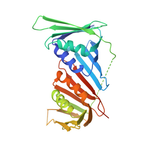
Structural analyses of PCNA from the fungal pathogen Candida albicans identify three regions with species-specific conformations.
Sundaram, R., Manohar, K., Patel, S.K., Acharya, N., Vasudevan, D.(2021) FEBS Lett 595: 1328-1349
- PubMed: 33544878
- DOI: https://doi.org/10.1002/1873-3468.14055
- Primary Citation of Related Structures:
7BUP - PubMed Abstract:
An assembly of multiprotein complexes achieves chromosomal DNA replication at the replication fork. In eukaryotes, proliferating cell nuclear antigen (PCNA) plays a vital role in the assembly of multiprotein complexes at the replication fork and is essential for cell viability. PCNA from several organisms, including Saccharomyces cerevisiae, has been structurally characterised. However, the structural analyses of PCNA from fungal pathogens are limited. Recently, we have reported that PCNA from the opportunistic fungal pathogen Candida albicans complements the essential functions of ScPCNA in S. cerevisiae. Still, it only partially rescues the loss of ScPCNA when the yeast cells are under genotoxic stress. To understand this further, herein, we have determined the crystal structure of CaPCNA and compared that with the existing structures of other fungal and human PCNA. Our comparative structural and in-solution small-angle X-ray scattering (SAXS) analyses reveal that CaPCNA forms a stable homotrimer, both in crystal and in solution. It displays noticeable structural alterations in the oligomerisation interface, P-loop and hydrophobic pocket regions, suggesting its differential function in a heterologous system and avenues for developing specific therapeutics. DATABASES: The PDB and SASBDB accession codes for CaPCNA are 7BUP and SASDHQ9, respectively.
Organizational Affiliation:
Laboratory of Macromolecular Crystallography, Department of Infectious Disease Biology, Institute of Life Sciences, Bhubaneswar, India.