7BPC

Crystal structure of 2, 3-dihydroxybenzoic acid decarboxylase from Fusarium oxysporum in complex with 2,5-DHBA


Experimental Data Snapshot

  • Method: X-RAY DIFFRACTION
  • Resolution: 2.45 Å
  • R-Value Free: 0.247 
  • R-Value Work: 0.193 
  • R-Value Observed: 0.196 

wwPDB Validation   3D Report Full Report


Ligand Structure Quality Assessment 


This is version 1.2 of the entry. See complete history


Literature

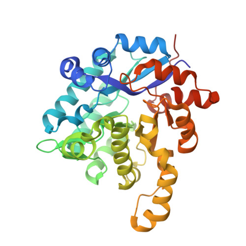
2,3-Dihydroxybenzoic Acid Decarboxylase from Fusarium oxysporum: Crystal Structures and Substrate Recognition Mechanism.

Song, M.Zhang, X.Liu, W.Feng, J.Cui, Y.Yao, P.Wang, M.Guo, R.T.Wu, Q.Zhu, D.

(2020) Chembiochem 21: 2950-2956

  • DOI: https://doi.org/10.1002/cbic.202000244
  • Primary Citation of Related Structures:  
    6M53, 7BP1, 7BPC

  • PubMed Abstract: 

    A 2,3-dihydroxybenzoic acid decarboxylase from Fusarium oxysporum (2,3-DHBD_Fo) has a relatively high catalytic efficiency for the decarboxylation of 2,3-dihydroxybenzoic acid (DHBA) and carboxylation of catechol, thus it has a different substrate spectrum from other benzoic acid decarboxylases. We have determined the structures of 2,3-DHBD_Fo in its apo form and complexes with catechol or 2,5-dihydroxybenzoic acid at 1.55, 1.97, and 2.45 Å resolution, respectively. The crystal structures of 2,3-DHBD_Fo show that the enzyme exists as a homotetramer, and each active center has a Zn 2+ ion coordinated by E8, H167, D291 and three water molecules. This is different from 2,6-DHBD from Rhizobium sporomusa, in which the Zn 2+ ion is also coordinated with H10. Surprisingly, mutation of A10 of 2,3-DHBD_Fo to His resulted in almost complete loss of the enzyme activity. Enzyme-substrate docking and site-directed mutation studies indicate that residue R233 Δ interacts with the 3-hydroxy group of 2,3-DHBA, and plays an important role in substrate recognition for this enzyme, thus revealing the molecular basis 2,3-dihydroxybenzoic acid decarboxylase.


  • Organizational Affiliation

    Key Laboratory of Industrial Fermentation Microbiology, Tianjin University of Science and Technology, Tianjin, 300457, P. R. China.


Macromolecules
Find similar proteins by:  (by identity cutoff)  |  3D Structure
Entity ID: 1
MoleculeChains Sequence LengthOrganismDetailsImage
2,3-dihydroxybenzoate decarboxylase
A, B, C, D
343Fusarium oxysporumMutation(s): 0 
Gene Names: BFJ70_g2310
UniProt
Find proteins for N1S495 (Fusarium oxysporum f. sp. cubense (strain race 4))
Explore N1S495 
Go to UniProtKB:  N1S495
Entity Groups  
Sequence Clusters30% Identity50% Identity70% Identity90% Identity95% Identity100% Identity
UniProt GroupN1S495
Sequence Annotations
Expand
  • Reference Sequence
Experimental Data & Validation

Experimental Data

  • Method: X-RAY DIFFRACTION
  • Resolution: 2.45 Å
  • R-Value Free: 0.247 
  • R-Value Work: 0.193 
  • R-Value Observed: 0.196 
  • Space Group: P 21 21 21
Unit Cell:
Length ( Å )Angle ( ˚ )
a = 76.769α = 90
b = 129.739β = 90
c = 140.557γ = 90
Software Package:
Software NamePurpose
PHENIXrefinement
HKL-2000data scaling
PDB_EXTRACTdata extraction
HKL-2000data reduction
PHASERphasing

Structure Validation

View Full Validation Report


Ligand Structure Quality Assessment 


Entry History 

Deposition Data

Revision History  (Full details and data files)

  • Version 1.0: 2020-07-15
    Type: Initial release
  • Version 1.1: 2020-10-28
    Changes: Database references
  • Version 1.2: 2023-11-29
    Changes: Data collection, Database references, Refinement description