7BOV

The Structure of Bacillus subtilis glycosyltransferase,Bs-YjiC


Experimental Data Snapshot

  • Method: X-RAY DIFFRACTION
  • Resolution: 2.29 Å
  • R-Value Free: 0.266 
  • R-Value Work: 0.234 
  • R-Value Observed: 0.237 

wwPDB Validation   3D Report Full Report


This is version 1.3 of the entry. See complete history


Literature

Structural and biochemical studies of the glycosyltransferase Bs-YjiC from Bacillus subtilis.

Liu, B.Zhao, C.Xiang, Q.Zhao, N.Luo, Y.Bao, R.

(2021) Int J Biol Macromol 166: 806-817

  • DOI: https://doi.org/10.1016/j.ijbiomac.2020.10.238
  • Primary Citation of Related Structures:  
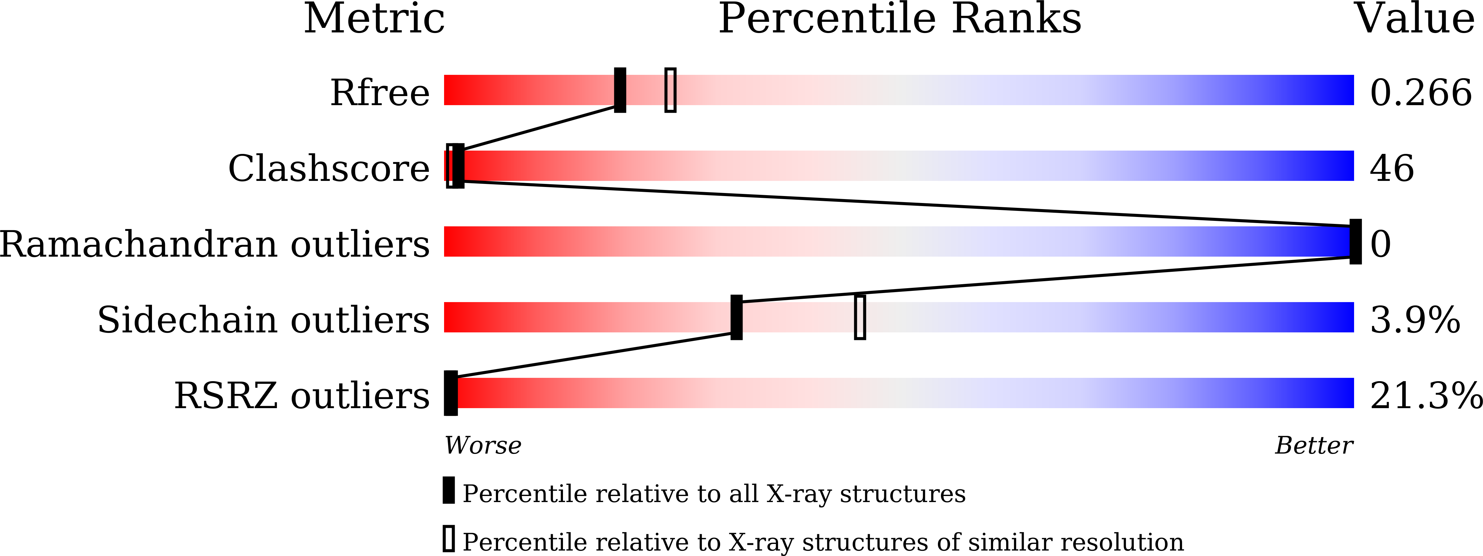
    7BOV

  • PubMed Abstract: 

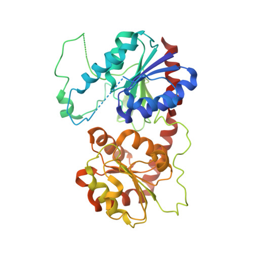
    Glycosylation possess prominent biological and pharmacological significance in natural product and drug candidate synthesis. The glycosyltransferase YjiC, discovered from Bacillus subtilis (Bs-YjiC), shows potential applications in drug development due to its wide substrate spectrums. In order to elucidate its catalytic mechanism, we solved the crystal structure of Bs-YjiC, demonstrating that Bs-YjiC adopts a typical GT-B fold consisting of a flexible N-domain and a relatively rigid C-domain. Structural analysis coupled with site-directed mutagenesis studies revealed that site Ser277 was critical for Nucleoside Diphosphate (NDP) recognition, while Glu317, Gln318, Ser128 and Ser129 were crucial for glycosyl moiety recognition. Our results illustrate the structural basis for acceptor promiscuity in Bs-YjiC and provide a starting point for further protein engineering of Bs-YjiC in industrial and pharmaceutical applications.


  • Organizational Affiliation

    Department of Gastroenterology, West China Hospital, Sichuan University and Collaborative Innovation Center of Biotherapy, Chengdu 610041, China.


Macromolecules
Find similar proteins by:  (by identity cutoff)  |  3D Structure
Entity ID: 1
MoleculeChains Sequence LengthOrganismDetailsImage
Uncharacterized UDP-glucosyltransferase YjiC392Bacillus subtilis subsp. subtilis str. 168Mutation(s): 0 
Gene Names: yjiCBSU12220
EC: 2.4.1
UniProt
Find proteins for O34539 (Bacillus subtilis (strain 168))
Explore O34539 
Go to UniProtKB:  O34539
Entity Groups  
Sequence Clusters30% Identity50% Identity70% Identity90% Identity95% Identity100% Identity
UniProt GroupO34539
Sequence Annotations
Expand
  • Reference Sequence
Experimental Data & Validation

Experimental Data

  • Method: X-RAY DIFFRACTION
  • Resolution: 2.29 Å
  • R-Value Free: 0.266 
  • R-Value Work: 0.234 
  • R-Value Observed: 0.237 
  • Space Group: I 2 2 2
Unit Cell:
Length ( Å )Angle ( ˚ )
a = 51.882α = 90
b = 118.433β = 90
c = 184.098γ = 90
Software Package:
Software NamePurpose
PHENIXrefinement
HKL-2000data reduction
HKL-2000data scaling
PHASERphasing

Structure Validation

View Full Validation Report


Entry History 

Deposition Data

Revision History  (Full details and data files)

  • Version 1.0: 2020-05-13
    Type: Initial release
  • Version 1.1: 2020-12-02
    Changes: Database references
  • Version 1.2: 2021-01-20
    Changes: Database references
  • Version 1.3: 2023-11-29
    Changes: Data collection, Database references, Refinement description