7AML

RET/GDNF/GFRa1 extracellular complex Cryo-EM structure


Experimental Data Snapshot

  • Method: ELECTRON MICROSCOPY
  • Resolution: 3.50 Å
  • Aggregation State: PARTICLE 
  • Reconstruction Method: SINGLE PARTICLE 

wwPDB Validation   3D Report Full Report


This is version 1.2 of the entry. See complete history


Literature

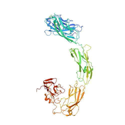
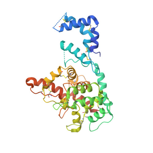
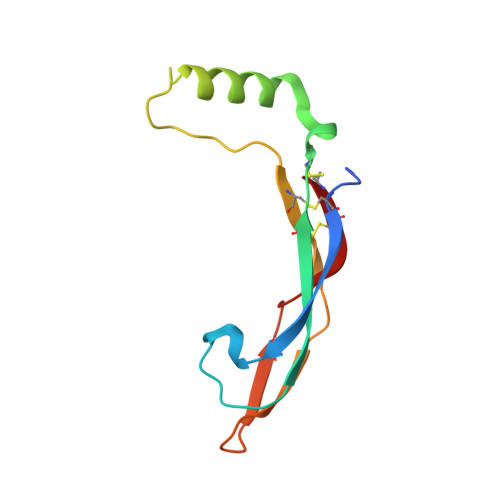
A two-site flexible clamp mechanism for RET-GDNF-GFR alpha 1 assembly reveals both conformational adaptation and strict geometric spacing.

Adams, S.E.Purkiss, A.G.Knowles, P.P.Nans, A.Briggs, D.C.Borg, A.Earl, C.P.Goodman, K.M.Nawrotek, A.Borg, A.J.McIntosh, P.B.Houghton, F.M.Kjaer, S.McDonald, N.Q.

(2021) Structure 29: 694

  • DOI: https://doi.org/10.1016/j.str.2020.12.012
  • Primary Citation of Related Structures:  
    7AB8, 7AMK, 7AML

  • PubMed Abstract: 

    RET receptor tyrosine kinase plays vital developmental and neuroprotective roles in metazoans. GDNF family ligands (GFLs) when bound to cognate GFRα co-receptors recognize and activate RET stimulating its cytoplasmic kinase function. The principles for RET ligand-co-receptor recognition are incompletely understood. Here, we report a crystal structure of the cadherin-like module (CLD1-4) from zebrafish RET revealing interdomain flexibility between CLD2 and CLD3. Comparison with a cryo-electron microscopy structure of a ligand-engaged zebrafish RET ECD -GDNF-GFRα1a complex indicates conformational changes within a clade-specific CLD3 loop adjacent to the co-receptor. Our observations indicate that RET is a molecular clamp with a flexible calcium-dependent arm that adapts to different GFRα co-receptors, while its rigid arm recognizes a GFL dimer to align both membrane-proximal cysteine-rich domains. We also visualize linear arrays of RET ECD -GDNF-GFRα1a suggesting that a conserved contact stabilizes higher-order species. Our study reveals that ligand-co-receptor recognition by RET involves both receptor plasticity and strict spacing of receptor dimers by GFL ligands.


  • Organizational Affiliation

    Signalling and Structural Biology Laboratory, Francis Crick Institute, NW1 1AT London, UK.


Macromolecules
Find similar proteins by:  (by identity cutoff)  |  3D Structure
Entity ID: 1
MoleculeChains Sequence LengthOrganismDetailsImage
Proto-oncogene tyrosine-protein kinase receptor RetA,
B [auth D]
615Danio rerioMutation(s): 0 
Gene Names: retret1
EC: 2.7.10.1
UniProt
Find proteins for A8E7C6 (Danio rerio)
Explore A8E7C6 
Go to UniProtKB:  A8E7C6
Entity Groups  
Sequence Clusters30% Identity50% Identity70% Identity90% Identity95% Identity100% Identity
UniProt GroupA8E7C6
Sequence Annotations
Expand
  • Reference Sequence
Find similar proteins by:  (by identity cutoff)  |  3D Structure
Entity ID: 2
MoleculeChains Sequence LengthOrganismDetailsImage
GDNF family receptor alphaC [auth B],
E
353Danio rerioMutation(s): 0 
Gene Names: gfra1agfralpha1a
UniProt
Find proteins for Q98TT9 (Danio rerio)
Explore Q98TT9 
Go to UniProtKB:  Q98TT9
Entity Groups  
Sequence Clusters30% Identity50% Identity70% Identity90% Identity95% Identity100% Identity
UniProt GroupQ98TT9
Sequence Annotations
Expand
  • Reference Sequence
Find similar proteins by:  (by identity cutoff)  |  3D Structure
Entity ID: 3
MoleculeChains Sequence LengthOrganismDetailsImage
Glial cell line-derived neurotrophic factorD [auth C],
F
101Danio rerioMutation(s): 0 
Gene Names: gdnf
UniProt
Find proteins for Q98TU0 (Danio rerio)
Explore Q98TU0 
Go to UniProtKB:  Q98TU0
Entity Groups  
Sequence Clusters30% Identity50% Identity70% Identity90% Identity95% Identity100% Identity
UniProt GroupQ98TU0
Sequence Annotations
Expand
  • Reference Sequence
Small Molecules
Ligands 2 Unique
IDChains Name / Formula / InChI Key2D Diagram3D Interactions
NAG
Query on NAG

Download Ideal Coordinates CCD File 
AA [auth D]
BA [auth D]
CA [auth C]
DA [auth F]
K [auth A]
AA [auth D],
BA [auth D],
CA [auth C],
DA [auth F],
K [auth A],
L [auth A],
M [auth A],
N [auth A],
O [auth A],
P [auth A],
Q [auth A],
V [auth D],
W [auth D],
X [auth D],
Y [auth D],
Z [auth D]
2-acetamido-2-deoxy-beta-D-glucopyranose
C8 H15 N O6
OVRNDRQMDRJTHS-FMDGEEDCSA-N
CA
Query on CA

Download Ideal Coordinates CCD File 
G [auth A]
H [auth A]
I [auth A]
J [auth A]
R [auth D]
G [auth A],
H [auth A],
I [auth A],
J [auth A],
R [auth D],
S [auth D],
T [auth D],
U [auth D]
CALCIUM ION
Ca
BHPQYMZQTOCNFJ-UHFFFAOYSA-N
Experimental Data & Validation

Experimental Data

  • Method: ELECTRON MICROSCOPY
  • Resolution: 3.50 Å
  • Aggregation State: PARTICLE 
  • Reconstruction Method: SINGLE PARTICLE 
EM Software:
TaskSoftware PackageVersion
RECONSTRUCTIONcryoSPARC2
MODEL REFINEMENTPHENIX1.18.2
MODEL REFINEMENTCoot

Structure Validation

View Full Validation Report


Entry History & Funding Information

Deposition Data


Funding OrganizationLocationGrant Number
The Francis Crick InstituteUnited Kingdom10115

Revision History  (Full details and data files)

  • Version 1.0: 2021-01-13
    Type: Initial release
  • Version 1.1: 2021-02-03
    Changes: Database references
  • Version 1.2: 2021-07-14
    Changes: Database references