7AL4

Ancestral Flavin-containing monooxygenase (FMO) 1 (mammalian)


Experimental Data Snapshot

  • Method: X-RAY DIFFRACTION
  • Resolution: 3.00 Å
  • R-Value Free: 0.264 
  • R-Value Work: 0.228 
  • R-Value Observed: 0.229 

wwPDB Validation   3D Report Full Report


Ligand Structure Quality Assessment 


This is version 1.2 of the entry. See complete history


Literature

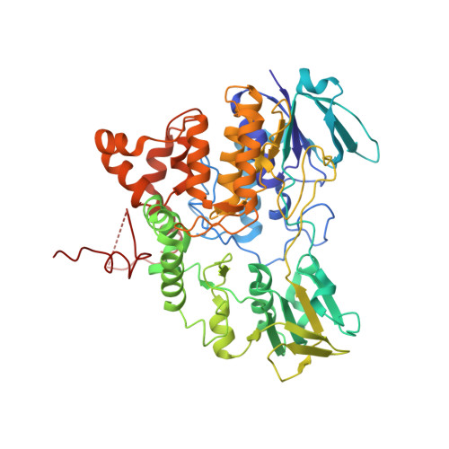
Ancestral reconstruction of mammalian FMO1 enables structural determination, revealing unique features that explain its catalytic properties.

Bailleul, G.Nicoll, C.R.Mascotti, M.L.Mattevi, A.Fraaije, M.W.

(2020) J Biol Chem 296: 100221-100221

  • DOI: https://doi.org/10.1074/jbc.RA120.016297
  • Primary Citation of Related Structures:  
    7AL4

  • PubMed Abstract: 

    Mammals rely on the oxidative flavin-containing monooxygenases (FMOs) to detoxify numerous and potentially deleterious xenobiotics; this activity extends to many drugs, giving FMOs high pharmacological relevance. However, our knowledge regarding these membrane-bound enzymes has been greatly impeded by the lack of structural information. We anticipated that ancestral-sequence reconstruction could help us identify protein sequences that are more amenable to structural analysis. As such, we hereby reconstructed the mammalian ancestral protein sequences of both FMO1 and FMO4, denoted as ancestral flavin-containing monooxygenase (AncFMO)1 and AncFMO4, respectively. AncFMO1, sharing 89.5% sequence identity with human FMO1, was successfully expressed as a functional enzyme. It displayed typical FMO activities as demonstrated by oxygenating benzydamine, tamoxifen, and thioanisole, drug-related compounds known to be also accepted by human FMO1, and both NADH and NADPH cofactors could act as electron donors, a feature only described for the FMO1 paralogs. AncFMO1 crystallized as a dimer and was structurally resolved at 3.0 Å resolution. The structure harbors typical FMO aspects with the flavin adenine dinucleotide and NAD(P)H binding domains and a C-terminal transmembrane helix. Intriguingly, AncFMO1 also contains some unique features, including a significantly porous and exposed active site, and NADPH adopting a new conformation with the 2'-phosphate being pushed inside the NADP + binding domain instead of being stretched out in the solvent. Overall, the ancestrally reconstructed mammalian AncFMO1 serves as the first structural model to corroborate and rationalize the catalytic properties of FMO1.


  • Organizational Affiliation

    Molecular Enzymology Group, University of Groningen, Groningen, the Netherlands.


Macromolecules
Find similar proteins by:  (by identity cutoff)  |  3D Structure
Entity ID: 1
MoleculeChains Sequence LengthOrganismDetailsImage
Ancestral Flavin-containing monooxygenase 1 (mammalian)A [auth D],
B [auth C],
C [auth B],
D [auth A]
531Felis catusMutation(s): 0 
Gene Names: FMO1
EC: 1.14.13.8
Entity Groups  
Sequence Clusters30% Identity50% Identity70% Identity90% Identity95% Identity100% Identity
Sequence Annotations
Expand
  • Reference Sequence
Small Molecules
Ligands 5 Unique
IDChains Name / Formula / InChI Key2D Diagram3D Interactions
FAD
Query on FAD

Download Ideal Coordinates CCD File 
E [auth D],
K [auth C],
M [auth B],
R [auth A]
FLAVIN-ADENINE DINUCLEOTIDE
C27 H33 N9 O15 P2
VWWQXMAJTJZDQX-UYBVJOGSSA-N
NAP
Query on NAP

Download Ideal Coordinates CCD File 
F [auth D],
L [auth C],
N [auth B],
S [auth A]
NADP NICOTINAMIDE-ADENINE-DINUCLEOTIDE PHOSPHATE
C21 H28 N7 O17 P3
XJLXINKUBYWONI-NNYOXOHSSA-N
LMT
Query on LMT

Download Ideal Coordinates CCD File 
G [auth D]
O [auth B]
P [auth B]
T [auth A]
V [auth A]
G [auth D],
O [auth B],
P [auth B],
T [auth A],
V [auth A],
W [auth A]
DODECYL-BETA-D-MALTOSIDE
C24 H46 O11
NLEBIOOXCVAHBD-QKMCSOCLSA-N
GOL
Query on GOL

Download Ideal Coordinates CCD File 
H [auth D],
J [auth D]
GLYCEROL
C3 H8 O3
PEDCQBHIVMGVHV-UHFFFAOYSA-N
CL
Query on CL

Download Ideal Coordinates CCD File 
I [auth D],
Q [auth B],
U [auth A]
CHLORIDE ION
Cl
VEXZGXHMUGYJMC-UHFFFAOYSA-M
Experimental Data & Validation

Experimental Data

  • Method: X-RAY DIFFRACTION
  • Resolution: 3.00 Å
  • R-Value Free: 0.264 
  • R-Value Work: 0.228 
  • R-Value Observed: 0.229 
  • Space Group: P 1 21 1
Unit Cell:
Length ( Å )Angle ( ˚ )
a = 115.919α = 90
b = 92.453β = 95.12
c = 156.688γ = 90
Software Package:
Software NamePurpose
REFMACrefinement
XDSdata reduction
Aimlessdata scaling
STARANISOdata scaling
PHASERphasing

Structure Validation

View Full Validation Report


Ligand Structure Quality Assessment 


Entry History & Funding Information

Deposition Data


Funding OrganizationLocationGrant Number
H2020 Marie Curie Actions of the European CommissionEuropean Union722390
H2020 Marie Curie Actions of the European CommissionEuropean Union847675

Revision History  (Full details and data files)

  • Version 1.0: 2020-12-30
    Type: Initial release
  • Version 1.1: 2021-04-07
    Changes: Database references
  • Version 1.2: 2024-01-31
    Changes: Data collection, Database references, Refinement description