Functional Characterization of the N -Acetylmuramyl-l-Alanine Amidase, Ami1, from Mycobacterium abscessus .
Kussau, T., Van Wyk, N., Johansen, M.D., Alsarraf, H.M.A.B., Neyret, A., Hamela, C., Sorensen, K.K., Thygesen, M.B., Beauvineau, C., Kremer, L., Blaise, M.(2020) Cells 9
- PubMed: 33158165
- DOI: https://doi.org/10.3390/cells9112410
- Primary Citation of Related Structures:
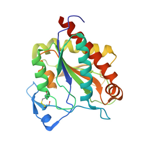
7AGL, 7AGM, 7AGO - PubMed Abstract:
Peptidoglycan (PG) is made of a polymer of disaccharides organized as a three-dimensional mesh-like network connected together by peptidic cross-links. PG is a dynamic structure that is essential for resistance to environmental stressors. Remodeling of PG occurs throughout the bacterial life cycle, particularly during bacterial division and separation into daughter cells. Numerous autolysins with various substrate specificities participate in PG remodeling. Expression of these enzymes must be tightly regulated, as an excess of hydrolytic activity can be detrimental for the bacteria. In non-tuberculous mycobacteria such as Mycobacterium abscessus , the function of PG-modifying enzymes has been poorly investigated. In this study, we characterized the function of the PG amidase, Ami1 from M. abscessus . An ami1 deletion mutant was generated and the phenotypes of the mutant were evaluated with respect to susceptibility to antibiotics and virulence in human macrophages and zebrafish. The capacity of purified Ami1 to hydrolyze muramyl-dipeptide was demonstrated in vitro. In addition, the screening of a 9200 compounds library led to the selection of three compounds inhibiting Ami1 in vitro. We also report the structural characterization of Ami1 which, combined with in silico docking studies, allows us to propose a mode of action for these inhibitors.
Organizational Affiliation:
Institut de Recherche en Infectiologie de Montpellier (IRIM), Université de Montpellier, CNRS UMR 9004, CEDEX 5, 34293 Montpellier, France.