7AD3

Class D GPCR Ste2 dimer coupled to two G proteins


Experimental Data Snapshot

  • Method: ELECTRON MICROSCOPY
  • Resolution: 3.50 Å
  • Resolution: 3.30 Å
  • Resolution: 3.30 Å
  • Resolution: 3.30 Å
  • Aggregation State: PARTICLE 
  • Reconstruction Method: SINGLE PARTICLE 

wwPDB Validation   3D Report Full Report


This is version 1.2 of the entry. See complete history


Literature

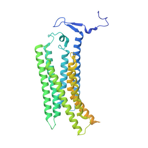
Structure of the class D GPCR Ste2 dimer coupled to two G proteins.

Velazhahan, V.Ma, N.Pandy-Szekeres, G.Kooistra, A.J.Lee, Y.Gloriam, D.E.Vaidehi, N.Tate, C.G.

(2021) Nature 589: 148-153

  • DOI: https://doi.org/10.1038/s41586-020-2994-1
  • Primary Citation of Related Structures:  
    7AD3

  • PubMed Abstract: 

    G-protein-coupled receptors (GPCRs) are divided phylogenetically into six classes 1,2 , denoted A to F. More than 370 structures of vertebrate GPCRs (belonging to classes A, B, C and F) have been determined, leading to a substantial understanding of their function 3 . By contrast, there are no structures of class D GPCRs, which are found exclusively in fungi where they regulate survival and reproduction. Here we determine the structure of a class D GPCR, the Saccharomyces cerevisiae pheromone receptor Ste2, in an active state coupled to the heterotrimeric G protein Gpa1-Ste4-Ste18. Ste2 was purified as a homodimer coupled to two G proteins. The dimer interface of Ste2 is formed by the N terminus, the transmembrane helices H1, H2 and H7, and the first extracellular loop ECL1. We establish a class D1 generic residue numbering system (CD1) to enable comparisons with orthologues and with other GPCR classes. The structure of Ste2 bears similarities in overall topology to class A GPCRs, but the transmembrane helix H4 is shifted by more than 20 Å and the G-protein-binding site is a shallow groove rather than a cleft. The structure provides a template for the design of novel drugs to target fungal GPCRs, which could be used to treat numerous intractable fungal diseases 4 .


  • Organizational Affiliation

    MRC Laboratory of Molecular Biology, Cambridge, UK.


Macromolecules
Find similar proteins by:  (by identity cutoff)  |  3D Structure
Entity ID: 1
MoleculeChains Sequence LengthOrganismDetailsImage
Pheromone alpha factor receptorA [auth B],
B [auth A]
431Saccharomyces cerevisiaeMutation(s): 0 
Gene Names: STE2
Membrane Entity: Yes 
UniProt
Find proteins for D6VTK4 (Saccharomyces cerevisiae (strain ATCC 204508 / S288c))
Explore D6VTK4 
Go to UniProtKB:  D6VTK4
Entity Groups  
Sequence Clusters30% Identity50% Identity70% Identity90% Identity95% Identity100% Identity
UniProt GroupD6VTK4
Sequence Annotations
Expand
  • Reference Sequence

Find similar proteins by:  Sequence   |   3D Structure  

Entity ID: 2
MoleculeChains Sequence LengthOrganismDetailsImage
Alpha-factor mating pheromoneC [auth K],
D [auth I]
13Saccharomyces cerevisiaeMutation(s): 0 
Membrane Entity: Yes 
UniProt
Find proteins for P01149 (Saccharomyces cerevisiae (strain ATCC 204508 / S288c))
Explore P01149 
Go to UniProtKB:  P01149
Entity Groups  
Sequence Clusters30% Identity50% Identity70% Identity90% Identity95% Identity100% Identity
UniProt GroupP01149
Sequence Annotations
Expand
  • Reference Sequence
Find similar proteins by:  (by identity cutoff)  |  3D Structure
Entity ID: 3
MoleculeChains Sequence LengthOrganismDetailsImage
STE4 isoform 1E [auth F]423Saccharomyces cerevisiaeMutation(s): 0 
Gene Names: STE4GI526_G0005548
UniProt
Find proteins for P18851 (Saccharomyces cerevisiae (strain ATCC 204508 / S288c))
Explore P18851 
Go to UniProtKB:  P18851
Entity Groups  
Sequence Clusters30% Identity50% Identity70% Identity90% Identity95% Identity100% Identity
UniProt GroupP18851
Sequence Annotations
Expand
  • Reference Sequence
Find similar proteins by:  (by identity cutoff)  |  3D Structure
Entity ID: 4
MoleculeChains Sequence LengthOrganismDetailsImage
Guanine nucleotide-binding protein alpha-1 subunit,Guanine nucleotide-binding protein alpha-1 subunitF [auth E],
H
233Saccharomyces cerevisiae S288CMutation(s): 6 
Gene Names: GPA1CDC70DAC1SCG1YHR005C
Membrane Entity: Yes 
UniProt
Find proteins for P08539 (Saccharomyces cerevisiae (strain ATCC 204508 / S288c))
Explore P08539 
Go to UniProtKB:  P08539
Entity Groups  
Sequence Clusters30% Identity50% Identity70% Identity90% Identity95% Identity100% Identity
UniProt GroupP08539
Sequence Annotations
Expand
  • Reference Sequence
Find similar proteins by:  (by identity cutoff)  |  3D Structure
Entity ID: 5
MoleculeChains Sequence LengthOrganismDetailsImage
Guanine nucleotide-binding protein subunit gamma109Saccharomyces cerevisiaeMutation(s): 2 
Gene Names: STE18GI526_G0003318PACBIOSEQ_LOCUS2168PACBIOSEQ_LOCUS3627PACBIOSEQ_LOCUS3692PACBIOSEQ_LOCUS3742PACBIOSEQ_LOCUS3767
Membrane Entity: Yes 
UniProt
Find proteins for P18852 (Saccharomyces cerevisiae (strain ATCC 204508 / S288c))
Explore P18852 
Go to UniProtKB:  P18852
Entity Groups  
Sequence Clusters30% Identity50% Identity70% Identity90% Identity95% Identity100% Identity
UniProt GroupP18852
Sequence Annotations
Expand
  • Reference Sequence
Experimental Data & Validation

Experimental Data

  • Method: ELECTRON MICROSCOPY
  • Resolution: 3.50 Å
  • Resolution: 3.30 Å
  • Resolution: 3.30 Å
  • Resolution: 3.30 Å
  • Aggregation State: PARTICLE 
  • Reconstruction Method: SINGLE PARTICLE 
EM Software:
TaskSoftware PackageVersion
RECONSTRUCTIONRELION3.1
RECONSTRUCTIONRELION
MODEL REFINEMENTPHENIX1.17.1-3660

Structure Validation

View Full Validation Report


Entry History & Funding Information

Deposition Data


Funding OrganizationLocationGrant Number
Medical Research Council (MRC, United Kingdom)United KingdomMRC U105197215
European Research Council (ERC)United KingdomEMPSI 339995

Revision History  (Full details and data files)

  • Version 1.0: 2020-12-09
    Type: Initial release
  • Version 1.1: 2020-12-16
    Changes: Database references
  • Version 1.2: 2021-01-13
    Changes: Database references