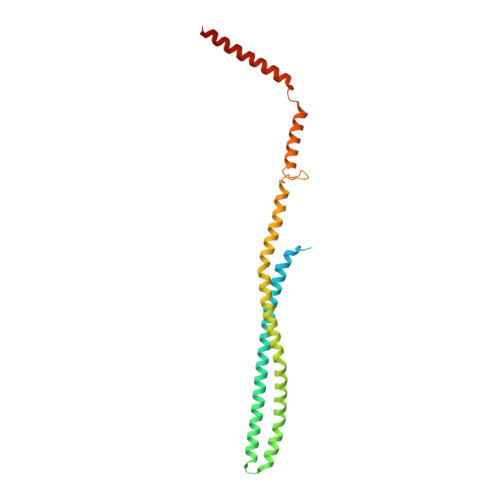
PspA adopts an ESCRT-III-like fold and remodels bacterial membranes.
Junglas, B., Huber, S.T., Heidler, T., Schlosser, L., Mann, D., Hennig, R., Clarke, M., Hellmann, N., Schneider, D., Sachse, C.(2021) Cell 184: 3674-3688.e18
- PubMed: 34166616
- DOI: https://doi.org/10.1016/j.cell.2021.05.042
- Primary Citation of Related Structures:
7ABK - PubMed Abstract:
PspA is the main effector of the phage shock protein (Psp) system and preserves the bacterial inner membrane integrity and function. Here, we present the 3.6 Å resolution cryoelectron microscopy (cryo-EM) structure of PspA assembled in helical rods. PspA monomers adopt a canonical ESCRT-III fold in an extended open conformation. PspA rods are capable of enclosing lipids and generating positive membrane curvature. Using cryo-EM, we visualized how PspA remodels membrane vesicles into μm-sized structures and how it mediates the formation of internalized vesicular structures. Hotspots of these activities are zones derived from PspA assemblies, serving as lipid transfer platforms and linking previously separated lipid structures. These membrane fusion and fission activities are in line with the described functional properties of bacterial PspA/IM30/LiaH proteins. Our structural and functional analyses reveal that bacterial PspA belongs to the evolutionary ancestry of ESCRT-III proteins involved in membrane remodeling.
Organizational Affiliation:
Ernst-Ruska Centre for Microscopy and Spectroscopy with Electrons, ER-C-3/Structural Biology, Forschungszentrum Jülich, 52425 Jülich, Germany; JuStruct: Jülich Center for Structural Biology, Forschungszentrum Jülich, 52425 Jülich, Germany; Department of Chemistry, Biochemistry, Johannes Gutenberg University Mainz, 55128 Mainz, Germany.