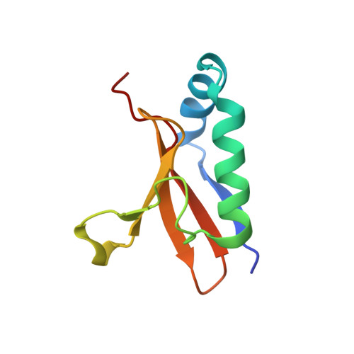
The two paralogous copies of the YoeB-YefM toxin-antitoxin module in Staphylococcus aureus differ in DNA binding and recognition patterns.
Xue, L., Khan, M.H., Yue, J., Zhu, Z., Niu, L.(2022) J Biol Chem 298: 101457-101457
- PubMed: 34861238
- DOI: https://doi.org/10.1016/j.jbc.2021.101457
- Primary Citation of Related Structures:
7V5Y, 7V5Z, 7V6W - PubMed Abstract:
Toxin-antitoxin (TA) systems are ubiquitous regulatory modules for bacterial growth and cell survival following stress. YefM-YoeB, the most prevalent type II TA system, is present in a variety of bacterial species. In Staphylococcus aureus, the YefM-YoeB system exists as two independent paralogous copies. Our previous research resolved crystal structures of the two oligomeric states (heterotetramer and heterohexamer-DNA ternary complex) of the first paralog as well as the molecular mechanism of transcriptional autoregulation of this module. However, structural details reflecting molecular diversity in both paralogs have been relatively unexplored. To understand the molecular mechanism of how Sa 2 YoeB and Sa 2 YefM regulate their own transcription and how each paralog functions independently, we solved a series of crystal structures of the Sa 2 YoeB-Sa 2 YefM. Our structural and biochemical data demonstrated that both paralogous copies adopt similar mechanisms of transcriptional autoregulation. In addition, structural analysis suggested that molecular diversity between the two paralogs might be reflected in the interaction profile of YefM and YoeB and the recognition pattern of promoter DNA by YefM. Interaction analysis revealed unique conformational and activating force effected by the interface between Sa 2 YoeB and Sa 2 YefM. In addition, the recognition pattern analysis demonstrated that residues Thr 7 and Tyr 14 of Sa 2 YefM specifically recognizes the flanking sequences (G and C) of the promoter DNA. Together, these results provide the structural insights into the molecular diversity and independent function of the paralogous copies of the YoeB-YefM TA system.
Organizational Affiliation:
Hefei National Laboratory for Physical Sciences at the Microscale, Division of Molecular and Cellular Biophysics, University of Science and Technology of China, Hefei, Anhui, China; Division of Life Sciences and Medicine, School of Life Sciences, University of Science and Technology of China, Hefei, Anhui, China.