


Crystal Structure of the Mitochondrial Ketol-acid Reductoisomerase IlvC from Candida auris
Kim, Y., Evdokimova, E., Di, R., Stogios, P., Savchenko, S., Joachimiak, A., Center for Structural Genomics of Infectious Diseases (CSGID)To be published.
Experimental Data Snapshot
Entity ID: 1 | |||||
|---|---|---|---|---|---|
| Molecule | Chains | Sequence Length | Organism | Details | Image |
| Ketol-acid reductoisomerase, mitochondrial | 363 | [Candida] auris | Mutation(s): 0 Gene Names: ilvC EC: 1.1.1.86 | 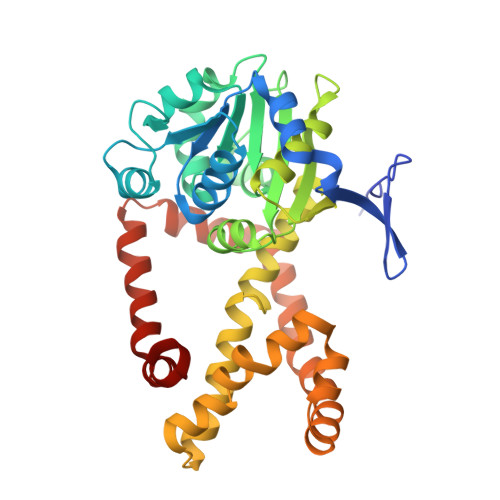 | |
UniProt | |||||
Find proteins for A0A2H0ZMH9 (Candida auris) Explore A0A2H0ZMH9 Go to UniProtKB: A0A2H0ZMH9 | |||||
Entity Groups | |||||
| Sequence Clusters | 30% Identity50% Identity70% Identity90% Identity95% Identity100% Identity | ||||
| UniProt Group | A0A2H0ZMH9 | ||||
Sequence AnnotationsExpand | |||||
| |||||
| Ligands 4 Unique | |||||
|---|---|---|---|---|---|
| ID | Chains | Name / Formula / InChI Key | 2D Diagram | 3D Interactions | |
| NDP (Subject of Investigation/LOI) Query on NDP | C [auth A], H [auth B] | NADPH DIHYDRO-NICOTINAMIDE-ADENINE-DINUCLEOTIDE PHOSPHATE C21 H30 N7 O17 P3 ACFIXJIJDZMPPO-NNYOXOHSSA-N |  | ||
| MLI Query on MLI | D [auth A], F [auth A] | MALONATE ION C3 H2 O4 OFOBLEOULBTSOW-UHFFFAOYSA-L |  | ||
| ACY Query on ACY | E [auth A], I [auth B] | ACETIC ACID C2 H4 O2 QTBSBXVTEAMEQO-UHFFFAOYSA-N |  | ||
| MG (Subject of Investigation/LOI) Query on MG | G [auth A], J [auth B], K [auth B], L [auth B] | MAGNESIUM ION Mg JLVVSXFLKOJNIY-UHFFFAOYSA-N |  | ||
| Length ( Å ) | Angle ( ˚ ) |
|---|---|
| a = 145.555 | α = 90 |
| b = 145.555 | β = 90 |
| c = 244.349 | γ = 120 |
| Software Name | Purpose |
|---|---|
| PHENIX | refinement |
| HKL-3000 | data reduction |
| HKL-3000 | data scaling |
| HKL-3000 | phasing |
| MOLREP | phasing |
| Funding Organization | Location | Grant Number |
|---|---|---|
| National Institutes of Health/National Institute Of Allergy and Infectious Diseases (NIH/NIAID) | United States | -- |
RCSB PDB (citation) is hosted by
RCSB PDB is a member of the

