


7P0H
Crystal structure of Helicobacter pylori ComF fused to an artificial alphaREP crystallization helper(named B2)
- PDB DOI: https://doi.org/10.2210/pdb7P0H/pdb
- Classification: RECOMBINATION
- Organism(s): synthetic construct, Helicobacter pylori 26695
- Expression System: Escherichia coli 'BL21-Gold(DE3)pLysS AG
- Mutation(s): No
- Deposited: 2021-06-29 Released: 2022-04-06
- Funding Organization(s): French Infrastructure for Integrated Structural Biology (FRISBI)
Experimental Data Snapshot
- Method: X-RAY DIFFRACTION
- Resolution: 2.50 Å
- R-Value Free: 0.245
- R-Value Work: 0.221
- R-Value Observed: 0.222

This is version 1.1 of the entry. See complete history.

Macromolecules
Find similar proteins by:
(by identity cutoff) | 3D Structure
Entity ID: 1 | |||||
|---|---|---|---|---|---|
| Molecule | Chains | Sequence Length | Organism | Details | Image |
| Helicobacter pylori ComF fused to an artificial alphaREP crystallization helper (named B2) | 427 | synthetic construct, Helicobacter pylori 26695 This entity is chimeric | Mutation(s): 0 | 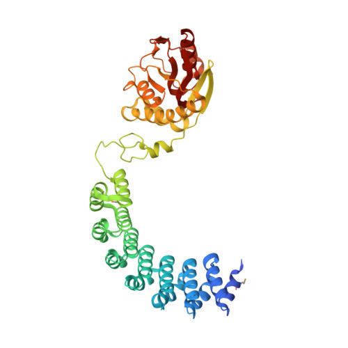 | |
UniProt | |||||
Find proteins for O26008 (Helicobacter pylori (strain ATCC 700392 / 26695)) Explore O26008 Go to UniProtKB: O26008 | |||||
Entity Groups | |||||
| Sequence Clusters | 30% Identity50% Identity70% Identity90% Identity95% Identity100% Identity | ||||
| UniProt Group | O26008 | ||||
Sequence AnnotationsExpand | |||||
| |||||
Small Molecules
| Ligands 4 Unique | |||||
|---|---|---|---|---|---|
| ID | Chains | Name / Formula / InChI Key | 2D Diagram | 3D Interactions | |
| PRP (Subject of Investigation/LOI) Query on PRP | DA [auth D], F [auth A], P [auth B], V [auth C] | 1-O-pyrophosphono-5-O-phosphono-alpha-D-ribofuranose C5 H13 O14 P3 PQGCEDQWHSBAJP-TXICZTDVSA-N |  | ||
| GOL Query on GOL | AA [auth C] FA [auth D] GA [auth D] H [auth A] I [auth A] | GLYCEROL C3 H8 O3 PEDCQBHIVMGVHV-UHFFFAOYSA-N |  | ||
| ZN (Subject of Investigation/LOI) Query on ZN | CA [auth D], E [auth A], O [auth B], U [auth C] | ZINC ION Zn PTFCDOFLOPIGGS-UHFFFAOYSA-N |  | ||
| MG (Subject of Investigation/LOI) Query on MG | BA [auth C] EA [auth D] G [auth A] HA [auth D] N [auth A] | MAGNESIUM ION Mg JLVVSXFLKOJNIY-UHFFFAOYSA-N |  | ||
| Modified Residues 1 Unique | |||||
|---|---|---|---|---|---|
| ID | Chains | Type | Formula | 2D Diagram | Parent |
| MSE Query on MSE | A, B, C, D | L-PEPTIDE LINKING | C5 H11 N O2 Se |  | MET |
Experimental Data & Validation
Experimental Data
- Method: X-RAY DIFFRACTION
- Resolution: 2.50 Å
- R-Value Free: 0.245
- R-Value Work: 0.221
- R-Value Observed: 0.222
- Space Group: P 1
Unit Cell:
| Length ( Å ) | Angle ( ˚ ) |
|---|---|
| a = 57.88 | α = 80.23 |
| b = 87.98 | β = 76.44 |
| c = 122.79 | γ = 76.39 |
| Software Name | Purpose |
|---|---|
| STARANISO | data scaling |
| BUSTER | refinement |
| PDB_EXTRACT | data extraction |
| XDS | data reduction |
| PHASER | phasing |
Entry History & Funding Information
Deposition Data
- Released Date: 2022-04-06 Deposition Author(s): Celma, L., Walbott, H., Legrand, P., Quevillon-Cheruel, S.
| Funding Organization | Location | Grant Number |
|---|---|---|
| French Infrastructure for Integrated Structural Biology (FRISBI) | France | ANR-10-INSB-05-01 |
Revision History (Full details and data files)
- Version 1.0: 2022-04-06
Type: Initial release - Version 1.1: 2022-04-27
Changes: Database references
