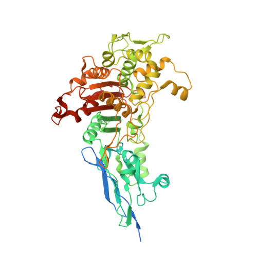
Structural Characterization of Diazabicyclooctane beta-Lactam "Enhancers" in Complex with Penicillin-Binding Proteins PBP2 and PBP3 of Pseudomonas aeruginosa.
Rajavel, M., Kumar, V., Nguyen, H., Wyatt, J., Marshall, S.H., Papp-Wallace, K.M., Deshpande, P., Bhavsar, S., Yeole, R., Bhagwat, S., Patel, M., Bonomo, R.A., van den Akker, F.(2021) mBio 12
- PubMed: 33593978
- DOI: https://doi.org/10.1128/mBio.03058-20
- Primary Citation of Related Structures:
7KIS, 7KIT, 7KIV, 7KIW - PubMed Abstract:
Multidrug-resistant (MDR) pathogens pose a significant public health threat. A major mechanism of resistance expressed by MDR pathogens is β-lactamase-mediated degradation of β-lactam antibiotics. The diazabicyclooctane (DBO) compounds zidebactam and WCK 5153, recognized as β-lactam "enhancers" due to inhibition of Pseudomonas aeruginosa penicillin-binding protein 2 (PBP2), are also class A and C β-lactamase inhibitors. To structurally probe their mode of PBP2 inhibition as well as investigate why P. aeruginosa PBP2 is less susceptible to inhibition by β-lactam antibiotics compared to the Escherichia coli PBP2, we determined the crystal structure of P. aeruginosa PBP2 in complex with WCK 5153. WCK 5153 forms an inhibitory covalent bond with the catalytic S327 of PBP2. The structure suggests a significant role for the diacylhydrazide moiety of WCK 5153 in interacting with the aspartate in the S-X-N/D PBP motif. Modeling of zidebactam in the active site of PBP2 reveals a similar binding mode. Both DBOs increase the melting temperature of PBP2, affirming their stabilizing interactions. To aid in the design of DBOs that can inhibit multiple PBPs, the ability of three DBOs to interact with P. aeruginosa PBP3 was explored crystallographically. Even though the DBOs show covalent binding to PBP3, they destabilized PBP3. Overall, the studies provide insights into zidebactam and WCK 5153 inhibition of PBP2 compared to their inhibition of PBP3 and the evolutionarily related KPC-2 β-lactamase. These molecular insights into the dual-target DBOs advance our knowledge regarding further DBO optimization efforts to develop novel potent β-lactamase-resistant, non-β-lactam PBP inhibitors. IMPORTANCE Antibiotic resistance is a significant clinical problem. Developing novel antibiotics that overcome known resistance mechanisms is highly desired. Diazabicyclooctane inhibitors such as zidebactam possess this potential as they readily inactivate penicillin-binding proteins, yet cannot be degraded by β-lactamases. In this study, we characterized the inhibition by diazabicyclooctanes of penicillin-binding proteins PBP2 and PBP3 from Pseudomonas aeruginosa using protein crystallography and biophysical analyses. These structures and analyses help define the antibiotic properties of these inhibitors, explain the decreased susceptibility of P. aeruginosa PBP2 to be inhibited by β-lactam antibiotics, and provide insights that could be used for further antibiotic development.
Organizational Affiliation:
Department of Biochemistry, Case Western Reserve University, Cleveland, Ohio, USA.