7KFY

Structural basis for a germline-biased antibody response to SARS-CoV-2 (RBD:C1A-F10 Fab)


Experimental Data Snapshot

  • Method: X-RAY DIFFRACTION
  • Resolution: 2.16 Å
  • R-Value Free: 0.249 
  • R-Value Work: 0.205 
  • R-Value Observed: 0.208 

wwPDB Validation   3D Report Full Report


Ligand Structure Quality Assessment 


This is version 1.2 of the entry. See complete history


Literature

Molecular basis for a germline-biased neutralizing antibody response to SARS-CoV-2.

Clark, S.A.Clark, L.E.Pan, J.Coscia, A.McKay, L.G.A.Shankar, S.Johnson, R.I.Griffiths, A.Abraham, J.

(2020) Biorxiv 

  • DOI: https://doi.org/10.1101/2020.11.13.381533
  • Primary Citation of Related Structures:  
    7KFV, 7KFW, 7KFX, 7KFY

  • PubMed Abstract: 

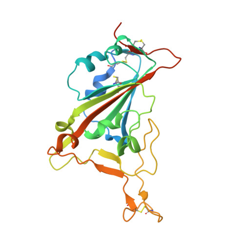
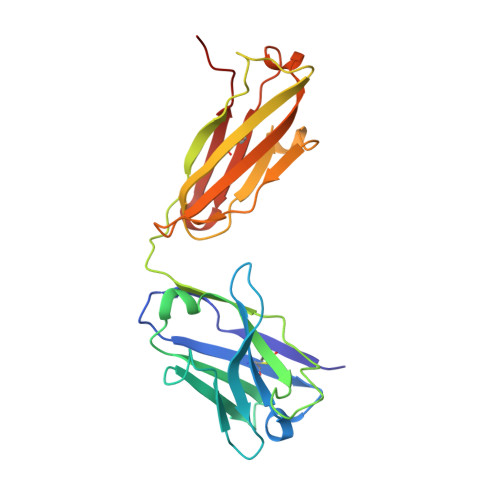
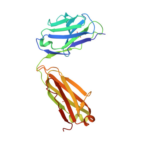
    The SARS-CoV-2 viral spike (S) protein mediates attachment and entry into host cells and is a major target of vaccine and drug design. Potent SARS-CoV-2 neutralizing antibodies derived from closely related antibody heavy chain genes (IGHV3-53 or 3-66) have been isolated from multiple COVID-19 convalescent individuals. These usually contain minimal somatic mutations and bind the S receptor-binding domain (RBD) to interfere with attachment to the cellular receptor angiotensin-converting enzyme 2 (ACE2). We used antigen-specific single B cell sorting to isolate S-reactive monoclonal antibodies from the blood of a COVID-19 convalescent individual. The seven most potent neutralizing antibodies were somatic variants of the same IGHV3-53-derived antibody and bind the RBD with varying affinity. We report X-ray crystal structures of four Fab variants bound to the RBD and use the structures to explain the basis for changes in RBD affinity. We show that a germline revertant antibody binds tightly to the SARS-CoV-2 RBD and neutralizes virus, and that gains in affinity for the RBD do not necessarily correlate with increased neutralization potency, suggesting that somatic mutation is not required to exert robust antiviral effect. Our studies clarify the molecular basis for a heavily germline-biased human antibody response to SARS-CoV-2.


Macromolecules
Find similar proteins by:  (by identity cutoff)  |  3D Structure
Entity ID: 1
MoleculeChains Sequence LengthOrganismDetailsImage
Spike glycoprotein228Severe acute respiratory syndrome coronavirus 2Mutation(s): 0 
Gene Names: S2
UniProt
Find proteins for P0DTC2 (Severe acute respiratory syndrome coronavirus 2)
Explore P0DTC2 
Go to UniProtKB:  P0DTC2
Entity Groups  
Sequence Clusters30% Identity50% Identity70% Identity90% Identity95% Identity100% Identity
UniProt GroupP0DTC2
Sequence Annotations
Expand
  • Reference Sequence
Find similar proteins by:  (by identity cutoff)  |  3D Structure
Entity ID: 2
MoleculeChains Sequence LengthOrganismDetailsImage
heavy chain of human antibody C1A-F10 FabB [auth H]225Homo sapiensMutation(s): 0 
Entity Groups  
Sequence Clusters30% Identity50% Identity70% Identity90% Identity95% Identity100% Identity
Sequence Annotations
Expand
  • Reference Sequence
Find similar proteins by:  (by identity cutoff)  |  3D Structure
Entity ID: 3
MoleculeChains Sequence LengthOrganismDetailsImage
light chain of human antibody C1A-F10 FabC [auth L]214Homo sapiensMutation(s): 0 
Entity Groups  
Sequence Clusters30% Identity50% Identity70% Identity90% Identity95% Identity100% Identity
Sequence Annotations
Expand
  • Reference Sequence
Small Molecules
Ligands 1 Unique
IDChains Name / Formula / InChI Key2D Diagram3D Interactions
NAG
Query on NAG

Download Ideal Coordinates CCD File 
D [auth A]2-acetamido-2-deoxy-beta-D-glucopyranose
C8 H15 N O6
OVRNDRQMDRJTHS-FMDGEEDCSA-N
Experimental Data & Validation

Experimental Data

  • Method: X-RAY DIFFRACTION
  • Resolution: 2.16 Å
  • R-Value Free: 0.249 
  • R-Value Work: 0.205 
  • R-Value Observed: 0.208 
  • Space Group: C 2 2 21
Unit Cell:
Length ( Å )Angle ( ˚ )
a = 85.74α = 90
b = 146.801β = 90
c = 144.593γ = 90
Software Package:
Software NamePurpose
BUSTERrefinement
PHENIXdata processing
Omodel building
XDSdata reduction
XDSdata scaling
PARROTphasing

Structure Validation

View Full Validation Report


Ligand Structure Quality Assessment 


Entry History & Funding Information

Deposition Data


Funding OrganizationLocationGrant Number
Other privateUnited States--

Revision History  (Full details and data files)

  • Version 1.0: 2020-12-02
    Type: Initial release
  • Version 1.1: 2021-01-20
    Changes: Database references, Source and taxonomy, Structure summary
  • Version 1.2: 2023-10-18
    Changes: Data collection, Database references, Refinement description