7JSY

Proteinase K soaked with I3C determined by MicroED from a single milled microcrystal

  • Classification: HYDROLASE
  • Organism(s): Parengyodontium album
  • Mutation(s): No 

  • Deposited: 2020-08-16 Released: 2020-10-14 
  • Deposition Author(s): Martynowycz, M.W., Gonen, T.
  • Funding Organization(s): Howard Hughes Medical Institute (HHMI), National Institutes of Health/National Institute of General Medical Sciences (NIH/NIGMS)

Experimental Data Snapshot

  • Method: ELECTRON CRYSTALLOGRAPHY
  • Resolution: 1.78 Å
  • R-Value Free: 0.209 
  • R-Value Work: 0.169 
  • R-Value Observed: 0.172 

wwPDB Validation   3D Report Full Report


This is version 1.1 of the entry. See complete history


Literature

Ligand Incorporation into Protein Microcrystals for MicroED by On-Grid Soaking.

Martynowycz, M.W.Gonen, T.

(2021) Structure 29: 88

  • DOI: https://doi.org/10.1016/j.str.2020.09.003
  • Primary Citation of Related Structures:  
    7JSY

  • PubMed Abstract: 

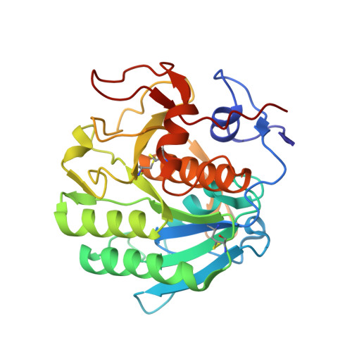
    A high throughout method for soaking ligands into protein microcrystals on TEM grids is presented. Every crystal on the grid is soaked simultaneously using only standard cryoelectron microscopy vitrification equipment. The method is demonstrated using proteinase K microcrystals soaked with the 5-amino-2,4,6-triodoisophthalic acid (I3C) magic triangle. A soaked microcrystal is milled to a thickness of approximately 200 nm using a focused ion beam, and MicroED data are collected. A high-resolution structure of the protein with four ligands at high occupancy is determined. Both the number of ligands bound and their occupancy is higher using on-grid soaking of microcrystals compared with much larger crystals treated similarly and investigated by X-ray crystallography. These results indicate that on-grid soaking ligands into microcrystals results in efficient uptake of ligands into protein microcrystals.


  • Organizational Affiliation

    Department of Biological Chemistry, University of California Los Angeles, 615 Charles E Young Drive South, Los Angeles, CA 90095, USA; Department of Physiology, University of California Los Angeles, 615 Charles E Young Drive South, Los Angeles, CA 90095, USA; Howard Hughes Medical Institute, University of California Los Angeles, Los Angeles CA90095, USA.


Macromolecules
Find similar proteins by:  (by identity cutoff)  |  3D Structure
Entity ID: 1
MoleculeChains Sequence LengthOrganismDetailsImage
Proteinase K279Parengyodontium albumMutation(s): 0 
EC: 3.4.21.64
UniProt
Find proteins for P06873 (Parengyodontium album)
Explore P06873 
Go to UniProtKB:  P06873
Entity Groups  
Sequence Clusters30% Identity50% Identity70% Identity90% Identity95% Identity100% Identity
UniProt GroupP06873
Sequence Annotations
Expand
  • Reference Sequence
Experimental Data & Validation

Experimental Data

  • Method: ELECTRON CRYSTALLOGRAPHY
  • Resolution: 1.78 Å
  • R-Value Free: 0.209 
  • R-Value Work: 0.169 
  • R-Value Observed: 0.172 
  • Space Group: P 43 21 2
Unit Cell:
Length ( Å )Angle ( ˚ )
a = 67.555α = 90
b = 67.555β = 90
c = 102.773γ = 90
Software Package:
Software NamePurpose
phenix.refinerefinement
PHENIXrefinement

Structure Validation

View Full Validation Report


Entry History & Funding Information

Deposition Data


Funding OrganizationLocationGrant Number
Howard Hughes Medical Institute (HHMI)United States--
National Institutes of Health/National Institute of General Medical Sciences (NIH/NIGMS)United StatesP41GM136508

Revision History  (Full details and data files)

  • Version 1.0: 2020-10-14
    Type: Initial release
  • Version 1.1: 2021-01-20
    Changes: Database references