7JMX

Crystal structure of a SARS-CoV-2 cross-neutralizing antibody COVA1-16 Fab


Experimental Data Snapshot

  • Method: X-RAY DIFFRACTION
  • Resolution: 2.53 Å
  • R-Value Free: 0.244 
  • R-Value Work: 0.211 
  • R-Value Observed: 0.213 

wwPDB Validation   3D Report Full Report


This is version 1.4 of the entry. See complete history


Literature

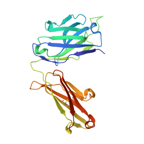
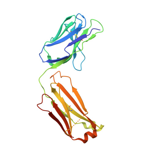
Cross-Neutralization of a SARS-CoV-2 Antibody to a Functionally Conserved Site Is Mediated by Avidity.

Liu, H.Wu, N.C.Yuan, M.Bangaru, S.Torres, J.L.Caniels, T.G.van Schooten, J.Zhu, X.Lee, C.D.Brouwer, P.J.M.van Gils, M.J.Sanders, R.W.Ward, A.B.Wilson, I.A.

(2020) Immunity 53: 1272-1280.e5

  • DOI: https://doi.org/10.1016/j.immuni.2020.10.023
  • Primary Citation of Related Structures:  
    7JMW, 7JMX

  • PubMed Abstract: 

    Most antibodies isolated from individuals with coronavirus disease 2019 (COVID-19) are specific to severe acute respiratory syndrome coronavirus 2 (SARS-CoV-2). However, COVA1-16 is a relatively rare antibody that also cross-neutralizes SARS-CoV. Here, we determined a crystal structure of the COVA1-16 antibody fragment (Fab) with the SARS-CoV-2 receptor-binding domain (RBD) and negative-stain electron microscopy reconstructions with the spike glycoprotein trimer to elucidate the structural basis of its cross-reactivity. COVA1-16 binds a highly conserved epitope on the SARS-CoV-2 RBD, mainly through a long complementarity-determining region (CDR) H3, and competes with the angiotensin-converting enzyme 2 (ACE2) receptor because of steric hindrance rather than epitope overlap. COVA1-16 binds to a flexible up conformation of the RBD on the spike and relies on antibody avidity for neutralization. These findings, along with the structural and functional rationale for epitope conservation, provide insights for development of more universal SARS-like coronavirus vaccines and therapies.


  • Organizational Affiliation

    Department of Integrative Structural and Computational Biology, The Scripps Research Institute, La Jolla, CA 92037, USA.


Macromolecules
Find similar proteins by:  (by identity cutoff)  |  3D Structure
Entity ID: 1
MoleculeChains Sequence LengthOrganismDetailsImage
COVA1-16 heavy chainA [auth H]238Homo sapiensMutation(s): 0 
Entity Groups  
Sequence Clusters30% Identity50% Identity70% Identity90% Identity95% Identity100% Identity
Sequence Annotations
Expand
  • Reference Sequence
Find similar proteins by:  (by identity cutoff)  |  3D Structure
Entity ID: 2
MoleculeChains Sequence LengthOrganismDetailsImage
COVA1-16 light chainB [auth L]214Homo sapiensMutation(s): 0 
Entity Groups  
Sequence Clusters30% Identity50% Identity70% Identity90% Identity95% Identity100% Identity
Sequence Annotations
Expand
  • Reference Sequence
Experimental Data & Validation

Experimental Data

  • Method: X-RAY DIFFRACTION
  • Resolution: 2.53 Å
  • R-Value Free: 0.244 
  • R-Value Work: 0.211 
  • R-Value Observed: 0.213 
  • Space Group: P 41 3 2
Unit Cell:
Length ( Å )Angle ( ˚ )
a = 156.287α = 90
b = 156.287β = 90
c = 156.287γ = 90
Software Package:
Software NamePurpose
MOLREPrefinement
PHENIXrefinement
HKL-2000data reduction
HKL-2000data scaling
PHASERphasing

Structure Validation

View Full Validation Report


Entry History & Funding Information

Deposition Data


Funding OrganizationLocationGrant Number
Bill & Melinda Gates FoundationUnited StatesOPP1170236
National Institutes of Health/National Institute Of Allergy and Infectious Diseases (NIH/NIAID)United StatesAI139445

Revision History  (Full details and data files)

  • Version 1.0: 2020-10-14
    Type: Initial release
  • Version 1.1: 2020-12-16
    Changes: Database references
  • Version 1.2: 2020-12-30
    Changes: Database references
  • Version 1.3: 2022-02-09
    Changes: Database references
  • Version 1.4: 2023-10-18
    Changes: Data collection, Database references, Refinement description